Influence of the Nervous System On Cardiac Output
Published .

Maintaining blood pressure is a complicated challenge that is handled very effectively by regulatory processes orchestrated primarily by the autonomic nervous system. These regulatory processes must operate over time scales ranging from very quick responses to changes in posture (e.g., a decrease in blood pressure when standing up) to responses to slower changes in blood pressure (e.g., a decrease in blood volume and blood pressure produced by dehydration).
Hypotension is a state of abnormally low blood pressure. Even a momentary state of hypotension that significantly decreases the delivery of blood to the brain can quickly produce dizziness or even a loss of consciousness (syncope), and this hypotensive state will be immediately apparent to the person experiencing it. Alternatively, an abnormally high blood pressure is a state of hypertension. Unlike hypotension, hypertension is not typically associated with any symptoms until it is extreme or very longstanding. Unfortunately, persistent hypertension can induce changes in the vasculature that increase the likelihood of stroke, damage the kidneys, and increase afterload that the heart must work against. By increasing afterload and making the heart work harder to force blood into the systemic circulation, hypertension can contribute to heart failure.
The moment-to-moment regulation of blood pressure is neurally mediated and requires three basic components:
- a sensory afferent component that includes pressure-sensitive nerve endings (baroreceptors) that are responsive to stretch of the artery wall as a proxy to monitor the pressure of arterial blood at specific locations in the cardiovascular system
- a “central control” that compares the “sensed pressure” with a desired pressure range
- efferent autonomic neural pathways from the CNS that can control heart rate, cardiac output, or systemic vascular resistance to maintain the sensed pressure close to the desired pressure
Baroreceptors
Baroreceptors are present at three locations in the cardiovascular system
- carotid sinuses
- aortic arch
- the atria of the heart
The baroreceptors in the carotid sinuses and aortic arch provide direct monitoring of the blood pressure in the systemic circulation, with the carotid sinuses providing monitoring closer to the point at which blood is delivered to the brain. Baroreceptors in the atria indirectly monitor blood volume via changes in central venous pressure.
Central Processing
The central processing of inputs from baroreceptors occurs in the hypothalamus and brainstem. Conceptually, we can imagine the sensory input from the baroreceptors being decoded into a representation of the blood pressure that can be compared with the desired blood pressure. Deviations from the desired blood pressure can then be corrected by adjustments to the efferent (i.e. neural output) pathways of the sympathetic and parasympathetic branches of the autonomic nervous system. These corrective responses for the sympathetic neural responses are examples of a negative feedback homeostatic mechanism .
Autonomic Control of the Cardiovascular System
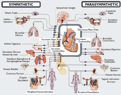
Most organs receive dual innervation from the parasympathetic and sympathetic branches of the autonomic nervous system.
The human heart typically has parasympathetic neural input that predominates in healthy, resting humans. The activity in the parasympathetic input to the sinoatrial node decreases the heart rate below the intrinsic rate of the node. Given the suppressive influence of this parasympathetic input, when an elevation in heart rate is needed, the first response is to reduce the activity of the parasympathetic input to the sinoatrial node, allowing the heart rate to rise passively towards the intrinsic rate. This withdrawal of parasympathetic activity is important during the oscillations in heart rate during respiration and during the initial phase of the rise in heart rate in response to low blood pressure upon standing, low grade exercise, and other minor perturbations. The effects of the parasympathetic input on the sino-atrial node are mediated by the release of the neurotransmitter acetylcholine from post-ganglionic parasympathetic fibers onto the target tissue.
When there is a need for an increase in heart rate, the activation of sympathetic inputs will raise the heart rate higher than its intrinsic rate to meet the increased demands. An increase in sympathetic nerve activity increases blood pressure by the following mechanisms:
- increasing heart rate, which increases cardiac output
- increasing stroke volume via increased contractility, which increases cardiac output
- constricting arterioles, which increases systemic vascular resistance
- constricting veins, which increases central venous pressure
- stimulating the release of renin from the kidney, leading to the production of angiotensin II and the release of aldosterone. Aldosterone stimulates the retention of sodium by the kidneys, which increases blood volume and venous return.
How does the sympathetic nervous system create these changes in the body? The effects of sympathetic nerve activity are mediated by the release of the neurotransmitter norepinephrine from post-ganglionic sympathetic fibers onto the target tissue. A similar effect on blood pressure can be produced by the stimulation by sympathetic preganglionic fibers of chromaffin cells in the adrenal medulla to release epinephrine (and norepinephrine) into the bloodstream. The circulating epinephrine (and norepinephrine) can activate the same receptors on the same target tissues as described above. There are two types of receptors for norepinephrine and epinephrine that are of particular interest at this point:
- ß1 (beta 1) receptors are present on the heart and on juxtaglomerular cells in the renal vasculature. The stimulation of ß1 receptors at the SA node increases the heart rate, and the stimulation of ß1 receptors on the contractile muscle of the heart increases the contractility (i.e., force of contraction). Both of these effects will increase cardiac output. The stimulation of ß1 receptors at the juxtaglomerular apparatus increases the release of renin, thereby increasing the production of angiotensin II. This produces an increase in blood volume and a brief period of vasoconstriction that increases systemic vascular resistance (SVR). All of these effects increase blood pressure because BP = CO x SVR. Therefore, we can lower blood pressure by administering drugs that block ß1 receptors. These are the “beta blocker” drugs.
- α1 (alpha 1) receptors are present on the smooth muscle that is present at arterioles. The stimulation of α1 receptors on arterioles causes contraction of the smooth muscle and constriction of the arteriole. A widespread constriction of arterioles increases systemic vascular resistance. Therefore, blood pressure can be lowered by drugs that block the activation of α1 receptors. These are the “alpha blocker” drugs.
