How People Die: Brain Misfire
Published .
Chemoreceptors
Adjustments of respiration and circulation in response to alterations in the levels of oxygen, carbon dioxide and hydrogen ions in the body fluids are mediated by two distinct chemoreceptive elements, situated peripherally and centrally. The peripheral arterial chemoreceptors, located in the carotid and aortic bodies, are supplied with sensory fibres coursing in the sinus and aortic nerves, and also receive sympathetic and parasympathetic motor innervations. The carotid receptors, and some aortic receptors, are essential for the immediate ventilatory and arterial pressure increases during acute hypoxic hypoxaemia, and also make an important contribution to respiratory compensation for acute disturbances of acid-base balance. The vascular effects of peripheral chemoreceptor stimulation include coronary vasodilation and vasoconstriction in skeletal muscle and the splanchnic area. The bradycardia and peripheral vasoconstriction during carotid chemoreceptor stimulation can be lessened or reversed by effects arising from a concurrent hyperpnoea. Central chemoreceptive elements respond to changes in the hydrogen ion concentration in the interstitial fluid in the brain, and are chiefly responsible for ventilatory and circulatory adjustments during hypercapnia and chronic disturbances of acid-base balance. The proposal that the neurones responsible for central chemoreception are located superficially in the ventrolateral portion of the medulla oblongata is not universally accepted, mainly because of a lack of convincing morphological and electrophysiological evidence. Central chemosensitive structures can modify peripheral chemoreceptor responses by altering discharges in parasympathetic and sympathetic nerves supplying these receptors, and such modifications could be a factor contributing to ventilatory unresponsiveness in mild hypoxia. Conversely, peripheral chemoreceptor drive can modulate central chemosensitivity during hypercapnia.
Stretch Receptors
Pulmonary stretch receptors are mechanoreceptors found in the lungs. When the lung expands, the receptors initiate the Hering-Breuer reflex, which reduces the respiratory rate. Increased firing from the stretch receptors also increases production of pulmonary surfactant. Intercostal muscles and diaphragm receive impulses from the respiratory center, stretch receptors in the lungs send impulses to the respiratory center giving information about the state of the lungs.
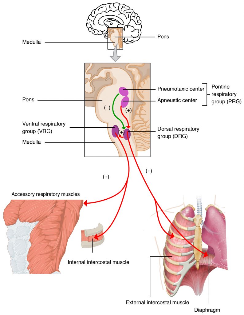
Medulla Rhythm Centers
The main respiratory muscles are under both voluntary and involuntary (automatic) control. These two control systems come from separate sites in the CNS and have separate descending pathways; the final integration of these outputs occurs at segmental levels in the cord. Voluntary control arises from the motor and premotor cortex and descends in the cord in the corticospinal tract. Involuntary control is mediated by both rhythmic and nonrhythmic systems located in the brainstem.
Respiration is controlled by the respiratory center in the brain stem in response to CO2 levels. Medulla Oblongata sets the basic rhythm of breathing (pacemaker). Pons smooths out respiratory rate and influence depth and length of respiration. A disturbance in the medulla or pons will interfere with the normal course of breathing (which is all about eliminating CO2) and result in hypoxia. Hypoxia, left unresolved will result in death.
The brain stem connects the brain to the spinal cord. This is essential for critical bodily functions, including:
- Heart regulation
- Blood circulation
- Heat regulation
- Breathing
- Swallowing, and
- Consciousness.
