Mechanics of Breathing
Published (updated: ).
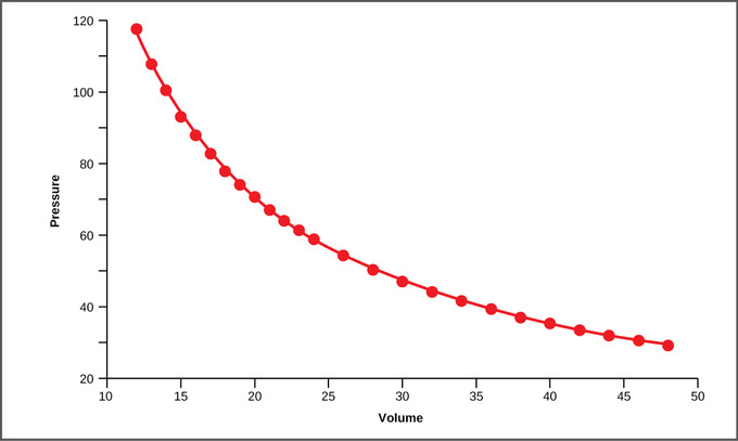
The relationship between gas pressure and volume helps to explain the mechanics of breathing. Boyle’s Law is the gas law which states that in a closed space, pressure and volume are inversely related. As volume decreases, pressure increases and vice versa. When discussing the detailed mechanics of breathing, it is important to keep this inverse relationship in mind.
The thoracic cavity, or chest cavity, always has a slight, negative pressure which aids in keeping the airways of the lungs open. During the process of inhalation, the lung volume expands as a result of the contraction of the diaphragm and intercostal muscles (the muscles that are connected to the rib cage), thus expanding the thoracic cavity. Due to this increase in volume, the pressure is decreased, based on the principles of Boyle’s Law. This decrease of pressure in the thoracic cavity relative to the environment makes the cavity pressure less than the atmospheric pressure. This pressure gradient between the atmosphere and the thoracic cavity allows air to rush into the lungs; inhalation occurs. The resulting increase in volume is largely attributed to an increase in alveolar space because the bronchioles and bronchi are stiff structures that do not change in size.
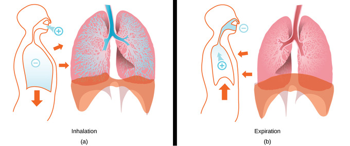
During this process, the chest wall expands out and away from the lungs. The lungs are elastic; therefore, when air fills the lungs, the elastic recoil within the tissues of the lung exerts pressure back toward the interior of the lungs. These outward and inward forces compete to inflate and deflate the lung with every breath. Upon exhalation, the lungs recoil to force the air out of the lungs. The intercostal muscles relax, returning the chest wall to its original position. During exhalation, the diaphragm also relaxes, moving higher into the thoracic cavity. This increases the pressure within the thoracic cavity relative to the environment. Air rushes out of the lungs due to the pressure gradient between the thoracic cavity and the atmosphere. This movement of air out of the lungs is classified as a passive event since there are no muscles contracting to expel the air.
Nervous and Muscular System Function In Breathing

The take home message in the following is that a patient requires an intact central nervous system to breath.
The respiratory center is located in the medulla oblongata and pons, in the brainstem. The respiratory center is made up of three major respiratory groups of neurons, two in the medulla and one in the pons. In the medulla they are the dorsal respiratory group, and the ventral respiratory group. In the pons, the pontine respiratory group includes two areas known as the pneumotaxic centre and the apneustic centre.
The respiratory center is responsible for generating and maintaining the rhythm of respiration, and also of adjusting this in homeostatic response to physiological changes. The respiratory center receives input from chemoreceptors, mechanoreceptors, the cerebral cortex, and the hypothalamus in order to regulate the rate and depth of breathing. Input is stimulated by altered levels of oxygen, carbon dioxide, and blood pH, by hormonal changes relating to stress and anxiety from the hypothalamus, and also by signals from the cerebral cortex to give a conscious control of respiration.
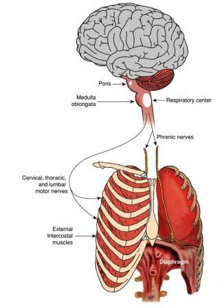
The phrenic nerve is a mixed motor/sensory nerve which originates from the C3-C5 spinal nerves in the neck. The nerve is important for breathing because it provides exclusive motor control of the diaphragm, the primary muscle of respiration. In humans, the right and left phrenic nerves are primarily supplied by the C4 spinal nerve, but there is also contribution from the C3 and C5 spinal nerves. From its origin in the neck, the nerve travels downward into the chest to pass between the heart and lungs towards the diaphragm.
The diaphragm is the primary muscle of respiration. The diaphragm, located below the lungs, is the major muscle of respiration. It is a large, dome-shaped muscle that contracts rhythmically and continually, and most of the time, involuntarily. Upon inhalation, the diaphragm contracts and flattens and the chest cavity enlarges. This contraction creates a vacuum, which pulls air into the lungs. Upon exhalation, the diaphragm relaxes and returns to its domelike shape, and air is forced out of the lungs.
The intercostal nerves emerge from the somatic nervous system and aid in the contraction of muscles as well as provide sensory information from the skin and parietal pleura. The intercostal muscles are several groups of muscles that run between the ribs, and help form and move the chest wall. The intercostal muscles are mainly involved in the mechanical aspect of breathing. These muscles help expand and shrink the size of the chest cavity to facilitate breathing
External intercostal muscles aid in quiet and forced inhalation. The external intercostals are responsible for the elevation of the ribs and bending them more open, thus expanding the transverse dimensions of the thoracic cavity.
