The Trachea and Bronchial Tree
Published (updated: ).
The Trachea
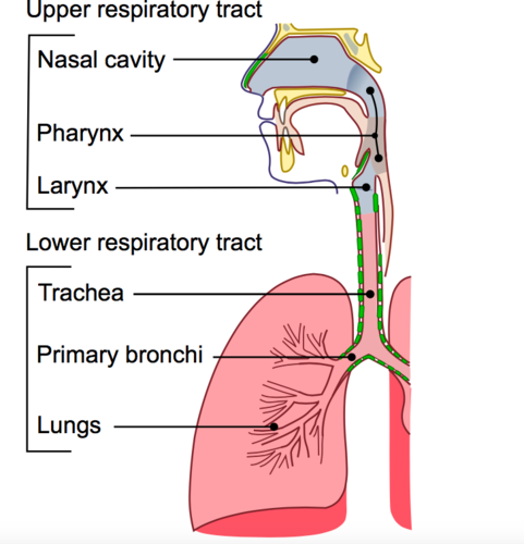
The larynx, commonly called the voice box or glottis, is the passageway for air between the pharynx above and the trachea below. It extends from the fourth to the sixth vertebral levels. The larynx is often divided into three sections: sublarynx, larynx, and supralarynx. It is formed by nine cartilages that are connected to each other by muscles and ligaments. The larynx plays an essential role in human speech. During sound production, the vocal cords close together and vibrate as air is expelled from the lungs and passes between them. The false vocal cords have no role in sound production, but help close off the larynx when food is swallowed.
The thyroid cartilage is the Adam’s apple. The epiglottis acts like a trap door to keep food and other particles from entering the larynx.
The trachea, commonly called the windpipe, is the main airway to the lungs. It divides into the right and left bronchi at the level of the fifth thoracic vertebra, channeling air to the right or left lung. The hyaline cartilage in the tracheal wall provides support and keeps the trachea from collapsing. The posterior soft tissue allows for expansion of the esophagus, which is immediately posterior to the trachea.
The mucous membrane that lines the trachea is ciliated pseudostratified columnar epithelium similar to that in the nasal cavity and nasopharynx. Goblet cells produce mucus that traps airborne particles and microorganisms, and the cilia propel the mucus upward, where it is either swallowed or expelled.
Bronchi and Bronchial Tree
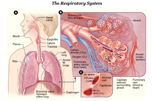
In the mediastinum (or space between the lungs), at the level of the fifth thoracic vertebra, the trachea divides into the right and left primary bronchi. The bronchi branch into smaller and smaller passageways until they terminate in tiny air sacs called alveoli.
The cartilage and mucous membrane of the primary bronchi are similar to that in the trachea. As the branching continues through the bronchial tree, the amount of hyaline cartilage in the walls decreases until it is absent in the smallest bronchioles. As the cartilage decreases, the amount of smooth muscle increases.
The alveolar ducts and alveoli consist primarily of simple squamous epithelium, which permits rapid diffusion of oxygen and carbon dioxide. Exchange of gases between the air in the lungs and the blood in the capillaries occurs across the walls of the alveolar ducts and alveoli.
The two lungs, which contain all the components of the bronchial tree beyond the primary bronchi, occupy most of the space in the thoracic cavity. The lungs are soft and spongy because they are mostly air spaces surrounded by the alveolar cells and elastic connective tissue. They are separated from each other by the mediastinum, which contains the heart. The only point of attachment for each lung is at the hilum, or root, on the medial side. This is where the bronchi, blood vessels, lymphatics, and nerves enter the lungs.
The right lung is shorter, broader, and has a greater volume than the left lung. It is divided into three lobes and each lobe is supplied by one of the secondary bronchi. The left lung is longer and narrower than the right lung. It has an indentation, called the cardiac notch, on its medial surface for the apex of the heart. The left lung has two lobes.
Each lung is enclosed by a double-layered serous membrane, called the pleura. The visceral pleura is firmly attached to the surface of the lung. At the hilum, the visceral pleura is continuous with the parietal pleura that lines the wall of the thorax. The small space between the visceral and parietal pleurae is the pleural cavity. It contains a thin film of serous fluid that is produced by the pleura. The fluid acts as a lubricant to reduce friction as the two layers slide against each other, and it helps to hold the two layers together as the lungs inflate and deflate.
