Respiratory System – More Structures
Published (updated: ).
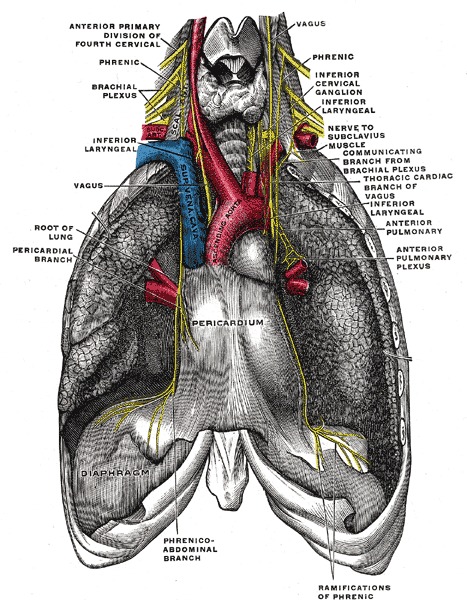
The phrenic nerve originates from the spinal cord at C3 through C5 and traverses the neck, heart, and lungs to reach the diaphragm and terminate into right and left branches at the subclavian artery.
The left phrenic nerve innervates the left diaphragmatic dome, and the right phrenic nerve innervates the right diaphragmatic dome, with the majority of nerve branching occurring on the inferior aspect of the diaphragm. The motor innervation activation will cause the diaphragm to contract with inspiration, resulting in a flattened diaphragm (the primary muscle of respiration) and increased intrapleural space. During exhalation, the diaphragm relaxes and returns to the dual dome shape. The phrenic nerve also provides touch and pain sensory innervation to the pericardium and intercostal nerves.
The phrenic nerve supplies sensory innervation to the diaphragm. Pain arising from the diaphragm is often referred to the tip of the shoulder, also known as the Kehr sign. For example, a patient with a subphrenic abscess or a ruptured spleen may complain of pain in the left shoulder. The hiccup reflex is due to irritation of the phrenic nerve. It results from sudden spasms of the diaphragm which pull air against the closed fold of the larynx. Once the phrenic nerve is injured, the diaphragm will become paralyzed.
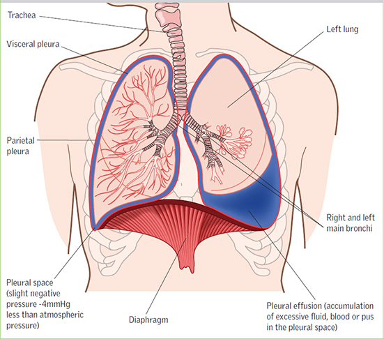
A pleura is a serous membrane that folds back on itself to form a two-layered membranous pleural sac. The outer layer is called the parietal pleura and attaches to the chest wall. The inner layer is called the visceral pleura and covers the lungs, blood vessels, nerves, and bronchi. There is no anatomical connection between the right and left pleural cavities. With the addition of pleural fluid, the lung pleura allows for easy movement of the lungs and inflation during breathing. The visceral pleura and parietal pleura connect via a vacuum lock called the pleural space. The parietal pleura are connect to the ribs and intercostal muscles. When contracted, the parietal pleura pull (via the vacuum lock) the lungs apart at all sides creating a vacuum.
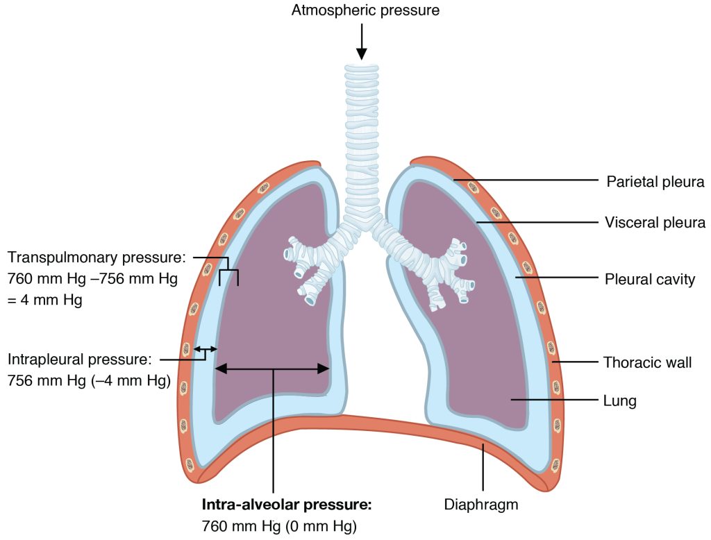
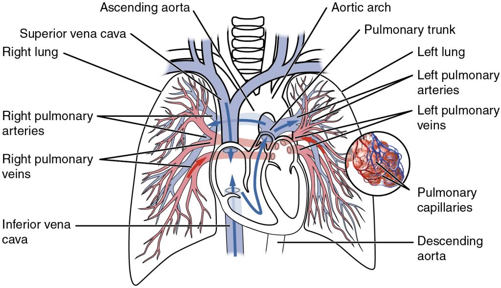
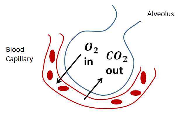
The left and right mainstem bronchi enter the lung through an airtight opening called the hilum. As the lungs are pulled in all different directions (primarily from the diaphragm), the air pressure inside the lungs become less than the environment (760 mmHg) creating a vacuum. Air is pulled through this negative pressure created by this vacuum travel through the pharynx, trachea, bronchi, bronchioles, and alveoli. Embedded on the alveoli is a complex network of pulmonary capillaries. The blood vessel walls of these tiny blood vessels are so tiny that oxygen and carbon dioxide can travel through the blood vessel walls.