Pediatric Trauma
Published (updated: ).
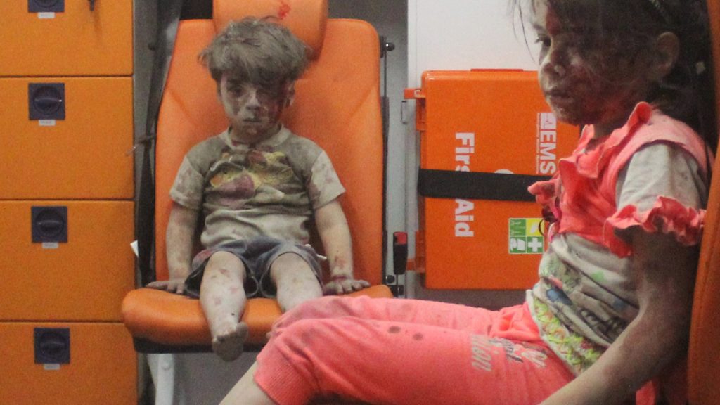
Anatomical, physiological, and psychological differences between children and adults have important implications for the initial assessment and management of pediatric trauma victims.
Children have less fat, more elastic connective tissue, and a pliable skeleton protecting tightly packed abdominal and thoracic structures. The force of an impact is transmitted widely through a child’s body, resulting in multisystem injuries in almost 50% of children with serious trauma. Their larger body surface area to body mass ratio predisposes them to larger heat and insensible fluid loss than adults, resulting in higher fluid and caloric requirements.
Children have a different physiological response to major trauma compared to adults, in that they maintain a near-normal blood pressure even in the face of 25% to 30% of blood volume loss. In these situations, subtle changes in the heart rate and extremity perfusion may signal impending cardiorespiratory failure, and should not be overlooked.
Children may not cope well emotionally in the aftermath of an accident. They need to be managed in a calm, child-friendly environment. The presence of a parent or guardian in the resuscitation room may help the trauma team by minimizing the injured child’s fear and anxieties. There is evidence that 25% of children suffer from post-traumatic stress disorder after a motor vehicle collision.
PRIMARY SURVEY
The primary survey is presented in a sequential fashion, but in reality the trauma team, directed by a team leader, performs the components of the primary survey simultaneously, so that the entire process takes only a few minutes. The goal of the primary survey is to find and relieve immediate life-threatening conditions. The primary survey starts at the injury scene and aims to ensure a patent airway, adequate breathing, circulatory support, and to assess major neurologic disability. The primary survey includes frequent reassessment to confirm or exclude injuries that require immediate surgical intervention.
AIRWAY
The assessment of the airway simply involves determining the ability of air to pass unobstructed into the lungs. The airway can be obstructed anywhere between the lips and the carina, by direct trauma, edema, secretions, blood, stomach contents, or foreign bodies. If the level of consciousness (LOC) is depressed, the child may not be able to maintain a patent airway and/or be able to protect the lungs from aspiration of stomach contents, because of loss of the gag reflex.
The classic sign of upper airway partial obstruction is inspiratory stridor. Respiratory effort with no air flow indicates complete airway obstruction.
BREATHING
Evaluate breathing to determine the child’s ability to ventilate and oxygenate.
Anticipate respiratory failure if any of the following signs is present:
- an increased respiratory rate, particularly with signs of distress (e.g. increased respiratory effort including nasal flaring, retractions, seesaw breathing, or grunting);
- an inadequate respiratory rate, effort, or chest excursion (e.g. diminished breath sounds or gasping), especially if mental status is depressed;
- cyanosis with abnormal breathing despite supplementary oxygen.
CERVICAL SPINE INJURIES
While spinal cord injuries are rare in children (<2% of injured children), a missed injury may have devastating consequences for the child, not to mention medico-legal ramifications for the trauma team. All significantly injured children must be assumed to have a cervical spine injury until proven otherwise by objective clinical examination. Approximately 30-40% of children with traumatic myelopathy have spinal cord injury without radiological abnormality.
DISABILITY
Perform a quick assessment of neurologic function at the end of the primary survey, and repeat during the secondary survey to monitor for changes in the child’s neurologic status. Causes of decreased level of consciousness in injured children include traumatic brain injury (TBI), hypoxemia, and poor cerebral perfusion. The latter two can exacerbate a TBI and result in secondary brain injury
INTRATHORACIC INJURIES
The vast majority of serious chest injuries in children are the result of blunt trauma. Most are the result of car and bicycle crashes. The presence of significant chest injury enhances the potential for multisystem trauma mortality by a factor of 10. Life-threatening thoracic injuries, such as airway obstruction, tension pneumothorax, massive hemothorax, and cardiac tamponade, are identified and treated during the primary survey.
The compliance of the child’s rib cage allows significant injury to occur with few obvious external signs of trauma. Energy is transmitted to the thoracic contents, and pulmonary contusions and hematomas are relatively more common, noted in more than 60% of children with severe thoracic injury. Because children have high oxygen consumption and low functional residual capacity, pulmonary contusions can result in severe hypoxemia, which may be refractory to oxygen therapy.
Commotio cordis is a disorder described in the pediatric population that results from a sudden impact to the anterior chest wall (such as a baseball injury) that causes cessation of normal cardiac function. The child may have an immediate dysrhythmia or ventricular fibrillation that is refractory to resuscitation efforts.
INTRA-ABDOMINAL INJURIES
Abdominal trauma is the most common cause of unrecognized fatal injury in children. Blunt trauma related to MVCs causes more than 50% of the abdominal injuries in children and is also the most lethal. Bicycle handlebars are a common cause of blunt abdominal trauma. Children have proportionally larger solid organs, less subcutaneous fat, and less protective abdominal musculature than adults. They suffer relatively more solid organ injury from both blunt and penetrating mechanisms. Approximately one-third of children with major trauma will have significant intra-peritoneal injuries.
AIRWAY MANAGEMENT INCLUDING C-SPINE STABILIZATION
If the airway is obstructed, inspect the mouth for a foreign body and remove it, but do not perform a blind finger sweep, which may push it further into the airway. Suction to clear blood, secretions, or vomitus. Perform an airway-opening maneuver: jaw thrust or chin lift. If there is any possibility of C-spine injury, do not perform a head tilt maneuver. If the child is unconscious, an oral airway may be required to lift the soft palate away from the base of the tongue. Bear in mind that inserting an oral airway into a semi-conscious child’s mouth may cause gagging and vomiting. Administer high-flow oxygen via a non-rebreathing face mask with an oxygen reservoir.
If the child is apneic or is making poor respiratory effort, assisted ventilation is required. When properly performed, bag-valve-mask (BVM) ventilation for a short period of time is as effective as ventilation via an ETT, and may be safer. A controlled trial of BVM versus ETT ventilation in an urban pre-hospital setting found no significant difference in survival or in the rate of achieving a good neurological outcome between the BVM group and the ETT group.
If an airway device has been placed prior to arrival in the trauma bay, the emergency department (ED) physician and/or anesthesiologist should not assume that it is the appropriate device, or that it has been correctly placed. The receiving team needs to be familiar with airway devices used by pre-hospital personnel, as they may differ from those used in the hospital. Capnography is the gold standard to confirm ventilation of the lungs. Capnography does not rule out mainstem bronchial intubation, however. Also, an ETT which is dislodged just proximal to the vocal cords could result in a waveform on the capnograph.
Presume that the child’s C-spine is injured until proven otherwise, especially in a child with a head injury. Techniques of immobilizing the C-spine include towel rolls, cervical collar, spinal board, and tape. Manual inline stabilization (MILS) of the C-spine is essential for intubation.
MANAGEMENT OF BREATHING, VENTILATION, AND OXYGENATION
For non-intubated patients arriving to the emergency room (ER), it is vital to assess, re-assess, and keep re-assessing ABCs for adequacy until the patient is transferred to the definitive care place (ICU or floor or the operating room). Pediatric victims of polytrauma have near-normal vital signs even in the presence of significant blood loss, and can deteriorate rapidly. These children should be monitored with extra vigilance during transport to the CT scanner, in the CT scanner, and in the emergency room.
Patients who arrive to the ER intubated should be monitored for existing or developing complications such as barotrauma or endobronchial intubation, in addition to ensuring that their oxygenation and ventilation is adequate.
If the child has confirmed or suspected head injury, and if ICP is monitored, mild hyperventilation may be needed for refractory increases in ICP. Prophylactic hyperventilation is not recommended, and may be harmful, by causing cerebral vasoconstriction and ischemia.
MANAGEMENT OF PAIN
Pain has, historically, been undertreated in patients presenting to the ED. This problem may be even greater in young pre-verbal injured children than in older children. Many physicians fear that administering opioids will mask symptoms of progressing injury. A recent meta-analysis found that the administration of opioids did not result in significant increase in management errors in patients presenting to the ED with abdominal pain.
If a regional anesthetic technique is not possible, a multimodal analgesic technique combining acetaminophen and NSAIDs reduces the dose of opioids required to treat pain. In the child who is NPO, IV acetaminophen and IV ketorolac can be given. IV patient-controlled analgesia (PCA) can be used in children above the age of about 5 years, and it allows the child to titrate opioid boluses according to the pain that they are experiencing.
PREVENTION OF HYPOTHERMIA
All victims of major trauma should be considered to be at risk for hypothermia. Children are more prone to develop hypothermia than adults. Hypothermia can lead to arrhythmias, coagulation abnormalities, and acidosis. The latter two, along with hypothermia, constitute “the triad of death” in trauma patients. An initial core temperature measurement (oral, rectal, or bladder) should be done as a part of the primary survey.
