Trauma In Pregnancy
Published (updated: ).
Introduction
Trauma in pregnancy has a wide spectrum, ranging from mild (single fall from standing height or striking the abdomen on an open drawer) to major (penetrating or high force blunt injury such as motor vehicle accident). Trauma in pregnancy has dramatically increased in the past 25 years and is now the number one cause of non-obstetrical maternal death in the United States. With major trauma there is a 40 to 50% risk of fetal death. Even with minor trauma, if it occurs during the first or second trimester there is an increase to delivering a child with prematurity or low birth weight. Although infrequently encountered in the clinical setting, emergency medicine physicians, trauma surgeons, and OBGYN’s should be aware of and prepared to manage a variety of complications associated with trauma in pregnancy. With sufficient knowledge of normal maternal physiology and potential pregnancy-related injury patterns, the physician facing a pregnant trauma victim will be better equipped to manage them, thus resulting in reduced morbidity and mortality.
Pathophysiology
Anatomic and Physiologic Changes in Pregnancy. To be able to recognize abnormal vital signs and injuries in a pregnant patient, normal anatomic and physiologic changes in pregnancy must be understood. Multiple organ systems undergo changes during pregnancy, the major, pertinent systems are discussed herein.
Abdominal Changes. The tone of the lower esophageal and gastric motility is reduced in pregnancy. This leads to reflux and retained food contents in the stomach, respectively. Because of these factors, there is an increased risk of aspiration, especially during intubation. The peritoneum stretches markedly and by the third trimester becomes much less sensitive to peritoneal irritation
Hematologic Changes. Both the plasma volume and the red cell mass increase throughout pregnancy. Plasma volume doubles by the end of the third trimester, however, in much higher proportion than the red blood cell mass increase. This results in dilutional anemia of pregnancy. The normal hemoglobin is 10-14 grams per deciliter by the term. The liver becomes hypermetabolic, increasing production of coagulation factors and fibrinogen. With this production, the patient is now more at risk for deep vein thrombosis and disseminated intravascular coagulation (DIC).
Pulmonary Changes. Respiratory rate (RR) x tidal volume (TV) = Minute ventilation (MV). RR does not change during pregnancy. Counterintuitively, TV is increased by 40%, leading to a 40% increase in MV. This leads to a lowered partial pressure of carbon dioxide (PCO) of 30 mmHg (normal values are in the range 35-45 mmHg), resulting in a chronically compensated respiratory alkalosis. This must be taken into account when evaluating a blood gas. The diaphragm is elevated by approximately 2-4 cm at term.
Cardiovascular Changes. Pregnancy-related cardiovascular changes require careful interpretation of the vital signs in the trauma patient. Pulse in the 3rd trimester elevates 15-20 beats per minute. The blood pressure goes down by 15-20 mmHg, however, returns to normal during the 3rd trimester. Any sign of hypotension should be evaluated immediately given this return to the normal level. It should not be attributed to the pregnant state as this is an interpretation of exclusion in the setting of trauma. Finally, as noted previously, the plasma volume rises by 50%, thus leading to a potentially delayed recognition of shock. By the time the maternal blood pressure falls, the patient may have already sustained a 30% blood loss.
Premature Labor. Premature labor is defined as the presence of uterine contractions occurring at less than 36 weeks’ gestation (premature) accompanied by cervical changes (labor). Although trauma patients may feel pain similar to real labor contractions, these pains may not represent contractions.
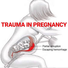
Placental Abruption. Placental abruption is the leading cause of fetal death not related to maternal death. It occurs in 1-5% of minor trauma. It is important to note that the classic triad of vaginal bleeding, abdominal pain, and uterine irritability may not be present. The edges of the placenta may encase the bleeding internally, in addition to the peritoneum being markedly insensitive given the massive stretching near term. Ultrasound may be helpful in the diagnosis but is not sensitive.
Amniotic Fluid Embolus. Uncommon and catastrophic, the pathophysiology of amniotic fluid embolus is currently poorly understood. The patient may present much like a massive pulmonary thrombotic embolus. There is no specific treatment for this entity, with exception of supportive care, intubation, vasopressors, and transfusion.
Uterine Rupture. Although a rare complication of trauma (0.06%), fetal mortality approaches 100% when present. It most often occurs in the 3rd trimester. Because of the high amount of force required to cause uterine rupture, it is commonly associated with pelvic fractures and bladder injuries. The classic presentation is dramatic; abdominal pain, distention (due to the unfolding of the fetus), palpable fetal parts, and shock. However, given the peritoneum’s insensitivity in the 3rd trimester, there may be no pain present. High index of suspicion accompanied by prompt recognition is critical. Treatment is exploratory laparotomy, delivery of fetus and supportive care.
History and Physical
Trauma assessment and resuscitation priorities are the same as in the nonpregnant woman but modified to thereafter address changes that occur in the later stages of pregnancy. This includes a primary, secondary, and tertiary survey. Attention should be paid to the vital signs as they change throughout normal pregnancy, noting changes to physiology as described previously. If alert and able, all women of child bearing age involved in trauma should be questioned on feasibility of pregnancy.
Clinical assessment alone may identify potential injury to the uterus or fetus. Concerning findings include: penetrating injury to the abdomen, vaginal bleeding, ruptured membranes, bulging perineum, presence of contractions and an abnormal fetal heart rate or rhythm. Vaginal bleeding is abnormal before labor and can be indicative of placental abruption, early labor, premature cervical dilation, or placentra previa. Rupture of the amniotic sac should be suspected when there is cloudy, white or green discharge, which leads to increased risk of infection. A ruptured amniotic sac may also lead to umbilical cord prolapse, which itself is an obstetric emergency requiring immediate cesarean section.
Evaluation
Fetal heart tones can be auscultated with a stethoscope at 20 weeks gestation; prior to this, a Doppler is needed. Evaluation of the fetus itself is best performed with toconometry, which is the most accurate means to detect fetal distress. Fetal heart tones can be detected as early as 12 weeks of gestation and are normally between 110 and 160 beats per minute (bpm). Initially, fetal hypoxia manifests as tachycardia, but as the arterial oxygen content decreases, the fetus ultimately becomes bradycardic. Therefore any sustained rate below 120 bpm should be recognized as fetal distress. If fetal distress is detected, then maternal blood loss must be suspected. Monitoring of fetal heart rate should be initiated on any woman greater than 24 weeks gestation, and is typically recommended for 4 to 6 hours duration after the initial presentation.
