Penetrating Head Trauma
Published (updated: ).
Penetrating brain injury (PBI) is a traumatic brain injury (TBI) which is a significant cause of mortality in young individuals. PBI includes all traumatic brain injuries other than blunt head trauma and constitutes the most severe of traumatic brain injuries.
Etiology
Based on the speed of penetration, it can be classified into two categories:
- High-velocity penetration: Examples include injuries caused by bullets or shell fragments, from direct trauma or shockwave injury to surrounding brain tissue due to a stretch injury.
- Low-velocity penetration: Examples include a knife or other sharp objects, with direct trauma to brain tissue.
Epidemiology
Traumatic brain injury (TBI) is one of the leading causes of death in the United States. Although penetrating trauma is less common than closed head trauma, it carries a worse prognosis. Most deaths from traumatic brain injury are due to firearm injuries. In the United States, approximately 20,000 gunshot injuries to the head occur annually.
Pathophysiology
The consequences of penetrating head injury depend on the following factors:
- Intracranial path and location: High mortality resulting from those that cross the midline, pass through the ventricles, or come to rest in the posterior fossa
- Energy and speed of entry: These factors depend on the properties of the weapon or missile. They result from energy being transferred from an object to the human skull and the underlying brain tissue. There is a high mortality rate associated with high-velocity projectiles. The kinetic energy involved is related to the square of the velocity. Three mechanisms of injuries have been reported.
- Laceration and crushing
- Cavitation
- Shockwaves
- Size and type of the penetrating object: Usually, large missiles or missiles that fragment within the cranial vault cause more fatalities
- Circumstances or events surrounding the injury
- Other associated injuries
Primary injuries occur immediately. Secondary injuries occur following the time of the injury. The final neurologic outcome is influenced by the extent and degree of secondary brain injury. Therefore, the primary goal in the emergency department is to prevent or reduce conditions that can worsen outcomes, such as hypotension, hypoxia, anemia, and hyperpyrexia.
The amount of damage to the brain depends on the kinetic energy imparted to the brain tissue. This, in turn, depends on the following factors:
- Trajectories of both the missile and the bone fragments through the brain
- Changes in intracranial pressure at the time of impact.
History and Physical
The presentation depends on the mechanism, site of the lesions, and associated injuries.
History should include:
- Date and time of injury
- Duration of loss of consciousness (LOC) if present
- Seizure at the time of impact
- Any co-morbidity (if existing)
- Anticoagulants and antiplatelet agents used
Initial physical examination includes primary and secondary trauma survey with the evaluation of other distracting injuries. A complete physical examination should be performed including a neurological examination. This should include documentation of the Glasgow coma scale (GCS). The involvement of cranial nerves should be assessed, and motor/sensory examination should be performed. It is important to realize that neurologic injury may be manifest distant to the site of impact. If unable to fully and formally assess cranial nerves secondary to lack of patient cooperation, it is important to, at least, document any findings relevant to the patient’s neurology.
Evaluation
In the pre-hospital setting, or in non-trauma facilities, stabilize, but, do not remove penetrating objects such as knives. Patients should be transported quickly to a location capable of providing definitive care. Early recognition of high-risk mechanisms, early imaging, and early evaluation at a level 1 trauma center may improve outcomes.
In the emergency department, resuscitation and stabilization should be provided. Perform a primary survey to identify any life-threatening injury. Stabilize, focusing on the airway, breathing, and circulation, including external hemorrhage, while establishing and maintaining cervical spine immobilization. Early activation of a trauma team may help to provide prompt recognition of polytrauma. The target is to maintain a systolic blood pressure of at least 90 mm Hg.
Following initial resuscitation and stabilization, an inspection of the superficial wound should be performed. Identify the entrance wound (and exit wounds, if present). Beware that blood-matted hair may cover these wounds. When a patient presents with a gunshot wound to the head, the other parts of the body including neck, chest, and abdomen should be inspected carefully for other gunshot wounds. Beware that injuries to the heart or great vessels in the chest or abdomen may be even more life-threatening.
Apply a sterile dressing to both the entrance and exit wounds. Assess whether there is any oozing of cerebrospinal fluid (CSF), blood, or brain parenchyma from the wound. Evaluate for hemotympanum, which may indicate a basilar skull fracture. Examine all orifices for retention of foreign bodies, the missile, teeth, and bone fragments.
Perform neurological examination, including GCS and document well. Evaluating for signs suggesting raised intracranial pressure is critical. The initial signs and symptoms may be nonspecific and include a headache, nausea, vomiting, and papilledema.
Perform a careful examination of the neck, chest, abdomen, pelvis, and extremities. Assume multiple injuries in cases of penetrating trauma. Obtain a detailed history including the “SAMPLE” history with an emphasis on events surrounding the injury. Also, determine the weapon type and/or caliber of the weapon.
Treatment / Management
Patients with penetrating head trauma require both medical and surgical management.
Medical Management
A low threshold for obtaining surgical consultation should be considered in cases of penetrating head trauma. Beware that many patients with penetrating head trauma will likely require operative intervention.
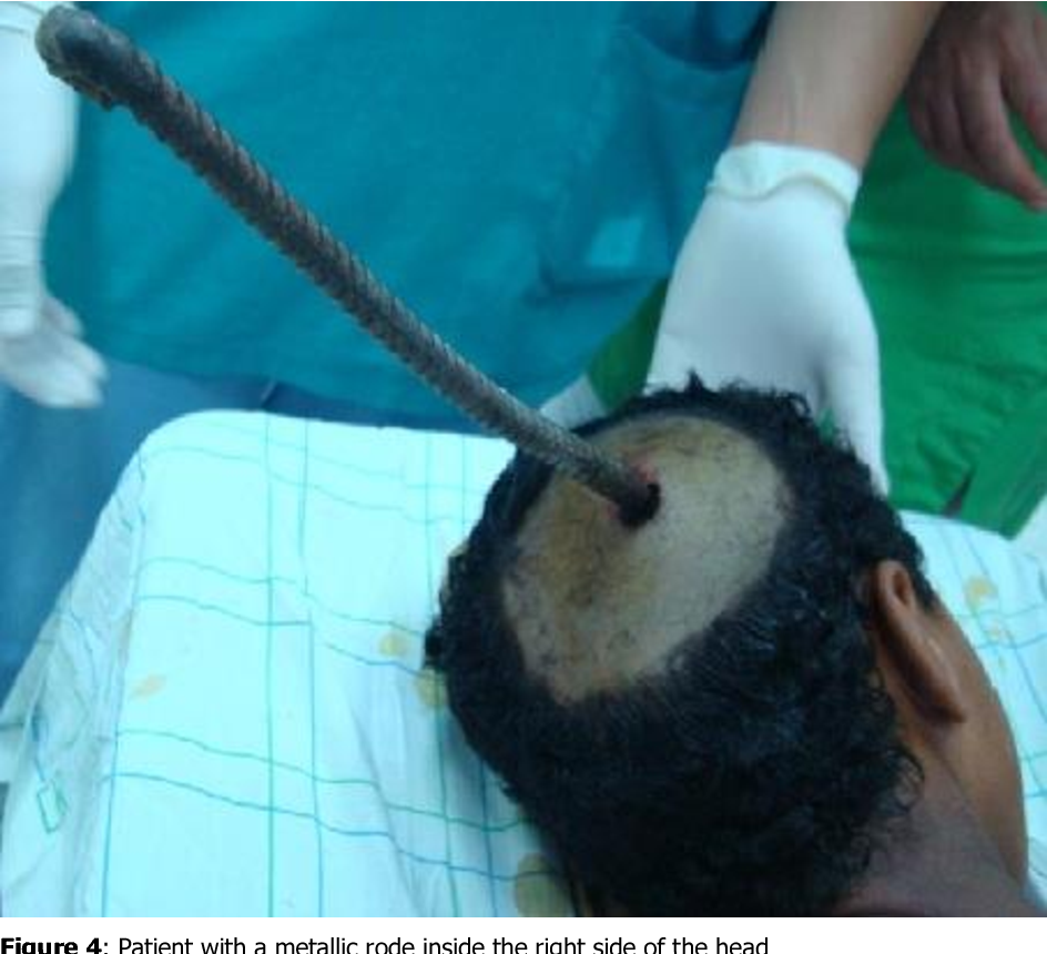
Indeed, do not remove any penetrating object from the skull in the emergency department until trauma and neurosurgical evaluation is obtained. Also, the protruding object should be stabilized, and provision should be made to protect it from moving during transportation of the patient, to prevent further injury.
Assess the need for endotracheal intubation.
- Inability to maintain adequate ventilation
- Inability to protect the airway due to depressed level of consciousness
- Neck or pharyngeal injury
Normalize PCO2. Avoid hyperventilation, because it leads to vasoconstriction and a subsequent reduction in the cerebral perfusion pressure (CPP). This may worsen long-term neurological outcome. Beware that hyperventilation is only a temporizing measure for the reduction of elevated intracranial pressure (ICP). Avoid hyperventilation during the first 24 hours after injury when cerebral blood flow (CBF) often is reduced.
