Tracheobronchial Tear
Published (updated: ).
Introduction
Tracheobronchial tear or laceration or injury is an uncommon injury to the tracheobronchial tree, usually involving the trachea or both the right and left main stem bronchi, and is associated with significant morbidity and mortality. Almost 80% of the tracheobronchial injuries in blunt trauma are expected to cause death on the site or during transport to the hospital, given multiple severe associated injuries. However, outcomes appear to be improving following better pre-hospital management. A high index of suspicion, early diagnosis, and management of tracheobronchial injuries are essential for a favorable outcome.
Etiology
The trachea is a relatively elastic structure made up of about 16 to 20 cartilaginous rings and is positioned and protected, given its vicinity to the sternum, rib cage, and vertebral column. The etiology for tracheobronchial tears includes penetrating and blunt trauma, and also iatrogenic injuries. Motor vehicle collisions represent more than half of the cases of penetrating and blunt traumas, causing tracheobronchial injury.
Penetrating injuries are more likely to cause a tracheobronchial tear than blunt trauma, and gunshot wounds are more common penetrating injuries than stab wounds. Blunt injuries are the less likely cause for tracheobronchial tear though the mortality is higher when compared with penetrating trauma.
Gunshot wounds generally cause injury to the cervical portion of the trachea, though any point of trachea could be injured. Stab injuries almost always involve the cervical part of the trachea. Blunt trauma to the cervical trachea could result indirectly from hyperextension injuries occurring while weightlifting or sudden deceleration during motor vehicle accidents. Direct cervical tracheal injuries result from wire strangulation while snow biking, seat belt, steering wheel, and dashboard injuries during the vehicular crash. Complex injuries involving both the above mechanisms could also happen in vehicular motor accidents as in “padded dashboard syndrome.” The thyroid and cricoid cartilages are most commonly involved in cervical tracheal injuries, and the area of disruption in severe injury is between the cricoid cartilage and trachea. Calcified laryngeal cartilages in adults are more likely to sustain fractures. In contrast, the flexible laryngeal cartilages are less likely injured in children when the entire brunt of trauma is targeted on the larynx.
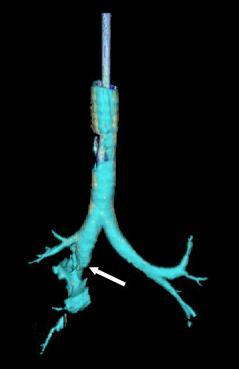
The lower thoracic trachea is often involved in blunt injuries, and the associated multiple severe injuries compound the higher mortality. Blunt trauma during motor vehicle accidents causes damage to the intrathoracic trachea, carina, and the main bronchi. The carina is the most fixed area in the airway system and hence is very likely affected by the shear stress forces during sudden deceleration. Thus blunt injuries are much more common in the lower trachea, which may vary from a partial horizontal tear to a complete separation. The vertical tear could go upwards from the carinal injury. The lower tracheal injury could extend to one or both bronchi resulting in a partial or complete laceration. Intra thoracic trachea usually gets injured with high energy trauma though low energy blows are also known to inflict injury. Most blunt bronchial injuries occur on the right side, likely due to the bulkier right lung and a shorter right main bronchus.
Pathophysiology
Penetrating injury most commonly involves the anterior trachea involving the cartilages or the ligamentous areas between the cartilages. Stab wounds are penetrating injuries almost always confined to the cervical trachea, whereas gunshot wounds could disrupt trachea or bronchi at any contact point. Blunt trauma causes tracheobronchial tears due to blow, shear stress, or burst mechanisms. Blunt trauma application of a large amount of energy to the anterior chest will push the lungs laterally & separates the bronchi from the relatively immobile carina. Carina is the most fixed part of the airway system in the chest and hence has an increased propensity to be inflicted by shear forces resulting from sudden deceleration. The sudden rise in airway pressure against closed glottis after a crush injury will result in the rupture of the airway whenever the pressure exceeds the tissue elasticity.
History and Physical
Though the location of airway tear will determine the presenting symptoms and signs, and also the optimal modality for identification, many patients present with non-specific symptoms irrespective of the site of injury. The common symptoms include breathing difficulty, stridor, and respiratory failure due to airway blockage, subcutaneous emphysema, hoarseness of voice or aphonia, hemoptysis, and other symptoms due to associated injury.
The most common presenting signs in tracheobronchial injury include subcutaneous emphysema (35% to 85%) and pneumothorax (20% to 50%). Hence the classic signs that have been described, which should trigger the suspicion of tracheobronchial injury in the appropriate settings, include subcutaneous emphysema, pneumomediastinum, and pneumothorax. Damage to proximal structures, including thoracic trachea and large bronchi, presents with pneumomediastinum with subcutaneous air, whereas more distal injury will result in pneumothorax and subcutaneous air. A raspy mediastinal crunch synchronizing with a heartbeat could be heard in mediastinal emphysema. Persistent air leak or non-resolving pneumothorax after an intercostal tube insertion should raise the suspicion of a tracheobronchial tear in a trauma setting.
