Pediatric Facial Fractures
Published (updated: ).

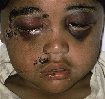
Trauma is a significant cause of morbidity and death in children. Trauma to the head is the most common anatomic site, and while facial fractures are infrequent, they can be severe and have life-long consequences. Younger patients have more elastic cartilaginous and bony structures in the face and a larger cranium: face ratio; thus, fractures in very young children are rare. The growth patterns of the face dictate age-specific fracture patterns that differ from adults. Though isolated facial fractures can occur, concurrent traumatic injuries to other nearby sites must always be considered, including injuries affecting the head, eyes, brain, neck, and airway.
Etiology
Facial fractures in children are typically caused by blunt trauma such as falls, sports injuries, transportation incidents (including automobiles, bicycles, skateboards, etc.), assault, and child abuse. Penetrating facial injuries, while rare, may also occur.
The neonatal skull is proportionally much larger than the face, and the forehead protrudes over the face. As the child grows, the face expands to comprise a greater relative area of the head until adult proportions are reached in the teenage years. Subsequently, traumatic head injuries in young children are more likely to spare the face while a skull injury is sustained.
Pediatric facial bones are more elastic and have more cartilage than adult bones; this allows more flexibility and compression. Given similar mechanisms to adult injuries, children have fewer relative facial fractures for this reason. When fractures occur, they are often minimally displaced and do not show the classic fracture patterns, such as Le Fort injuries described in adults. Nasal fractures are the most common fracture overall due to the prominence of the nasal bridge and minimal surrounding structural support.
Fracture sites relate to age-dependent development of the sinuses and, to a lesser degree, the stage of dentition. During development, bone initially thickens before becoming fully pneumatized and thinning into the final adult structural bone. The site of active bone growth and early pneumatization will be thicker and more resistant to fractures than later thin adult-like bone.
Epidemiology
Each year in the United States, pediatric trauma causes approximately 12,000 deaths, and at least 8 million ER encounters. Among all patients with facial fractures, fewer than 15% are children. Most facial injuries in children are limited to soft tissues, with only 10 to 15% of pediatric facial injuries resulting in facial fractures. However, more than half of all facial trauma presentations are associated with concurrent additional severe injuries beyond the face.
Many minor facial traumas are treated at home and maybe underreported; fractures are likely to cause significant pain and swelling and are, therefore, most likely more accurately reported than soft tissue trauma to the face. Males are more likely to have facial fractures than females, especially during adolescence, when males are approximately twice as likely to present with fractures than females.
Facial fractures are rare below the age of six, wherein skull fractures are more likely to be the result of head or facial trauma. Half of all fracture presentations are seen in ages 10 to 18 years. When fractures occur, roughly half are the result of motor vehicle collisions. Beyond motor vehicle collisions, bicycle accidents and sports injuries comprise most of the remaining trauma etiologies in school-age children, while infants and toddlers are more likely to suffer from falls. Adolescent males are the demographic group most likely to become injured from assault.
Fracture location can be age-dependent, both due to activities undertaken affecting the location of likely blunt trauma and differential areas of bone growth and laxity with age. Nasal fractures are generally thought to be the most common facial fractures, though they are likely underreported as they do not necessitate evaluation at a trauma center from which most pediatric facial trauma data are drawn.
Fracture patterns described in adults, such as Le Fort injuries, are rare in pediatric patients. The changes made through facial development correspond to different stress points for fracture locations; these patterns are generally only seen in older adolescents and account for less than 2% of fractures.
History and Physical
History
An accurate account of the events in a traumatic injury, including changes in mental status, sensory and motor function, range of motion, vision, and associated symptoms, is critical in evaluating trauma. Considering the child’s age, supporting history from parents, coaches, and first responders are likely needed. Children rarely have underlying medical conditions that contribute to a traumatic presentation or offer complications such as the use of anticoagulation; nonetheless, these routine historical questions should still be pursued as they may change management, along with reports of allergies, vaccinations (particularly tetanus), and last meal.
Patients might report swelling or stiffness in the head, neck, jaw, eyes, or nose. Feeling that teeth are loose, or the presence of or history of epistaxis does not change the likelihood of facial fractures. Sensations that something is stuck or catching, persistent diplopia, subjective malocclusion, or paresthesias of the face should raise concern for fractures.
Physical Exam
A careful physical exam is critical in children. Depending on age, they may not communicate a complete history of the traumatic event or relay all of their symptoms.
A calm, relaxed patient will be more accommodating to an extensive and careful examination. Examination success can be improved by being held by a parent and consideration of pain control, distraction, and anxiolysis.
A facial examination should be systematic. The exact approach is not as important as long as all aspects are examined. One method is to attempt the exam in 3 dimensions: superior to inferior, lateral to medial, then superficial to deep.
Musculoskeletal and Skin
For wound evaluations, determine the depth and explore any damage to muscles, tendons, vessels, nerves, and ducts. Facial nerve palsy after blunt trauma is suspicious for fracture of the temporal bone. Some amount of pain and stiffness with the range of motion is expected after trauma. Bony tenderness and soft tissue swelling are suggestive of, but nonspecific for, facial bone fractures; however, crepitus near a sinus more strongly correlates with an underlying fracture.
Eyes
If direct eye trauma has occurred, an examination of the eye should occur early as periorbital swelling can develop and hinder a later exam. An extraocular range of motion impairment suggests entrapment, possibly in a fracture of the orbital rim.
Mouth and Intraoral Examination
The examination of the mouth for fractures focuses on the upper and lower jaws, teeth, and temporomandibular joint (TMJ). In addition to bony tenderness, particularly trismus, malocclusion, or dental laxity with palpation, gingival ecchymosis, or lacerations may be signs of fractures in the mandible or maxilla.
Treatment / Management
As with most traumatic injuries, pediatric facial fractures benefit from ice, rest, and pain control. Once fractures are identified, the appropriate specialists should be consulted for further management and treatment recommendations. These specialists may include any of the following: pediatric specialists in facial surgery (both otolaryngology and plastic surgery, depending on local resources), ophthalmology, neurosurgery, anesthesia (for advanced airway stabilization), psychiatry (if self-harm is suspected), as well as any other team that is clinically indicated for consultation.
Prognosis
Pediatric facial trauma prognosis is generally good, though the more bones involved adds to the chance for long term deformity and need for surgical repair. Reassuringly, pediatric osteochondral tissues are adept at remodeling, and most patients heal well with minimal later discernable evidence of injury.
Complications
Possible complications include:
- Growth abnormality or long term disfiguration
- Infection (especially if dental or sinus involvement, or penetration of foreign objects)
- Long term dental effects
- Psycho-social effects
- Visual acuity distortion (especially for delayed relief of entrapped orbital muscles)
- Persistent paresthesia or muscle weakness if peripheral nerves damaged
- Posttraumatic or chronic facial pain
