Intraosseous Infusion
Published .
Introduction
Intraosseous (IO) vascular access refers to the placement of a specialized hollow bore needle through the cortex of a bone into the medullary space for infusion of medical therapy and laboratory tests. The IO route is an option when standard venous access would delay therapy or is not easily obtained in the hospital or pre-hospital setting.
IO success rates are twice as high as intravenous line placement in critical trauma patients without a blood pressure and should have priority over IV placement. IO needle insertion has been shown by multiple studies to have high success rates by physicians, nurses, and paramedics in adults, pediatrics patients, animals, as well as sim model studies.
Although IO access is superior in many clinical situations, it is highly underutilized. Studies show that IO access can be acquired within 20 seconds, allowing rapid access in emergent patients who would otherwise be challenging to access intravenously. Despite the proven value of IO access in the critical patient, barriers exist to its use. These barriers include a lack of confidence in the indications for using IO access by physicians and the belief that nursing staff is not familiar with IO access.
IO can be used to administer any substance that is infusible intravenously, but IO use should not be for longer than 24 hours due to an increased risk of complications.
Multiple IO devices are available from manufacturers, and availability varies institutionally. IO is easier than standard venous access and central lines in many situations and acceptable for all age groups, including preterm neonates.
Anatomy and Physiology
Sternum, clavicle, humeral head, iliac crest, distal femur, proximal tibia, distal tibia, and calcaneus are all potential sites for intraosseous access. The proximal tibia, humeral head, and sternum are the preferred sites in adults. The distal femur, proximal tibia, and distal tibia are preferred sites for infants and neonates. Always palpate both margins of the boney site to ensure penetration of the bone centrally. Note that each site is always one to two fingerbreadths in measurement to locate the correct location.
- Sternum: 1 cm below the sternal notch.
- Humerus: The humerus should be internally rotated and the hand placed on the abdomen with the elbow flexed to 90 degrees, ensuring that the bicep tendon is medially located and not penetrated. The surgical neck is palpated, and the needle is placed 2 cm above the surgical neck into the greater tubercle at about 45 degrees to the anterior plane. A longer IO needle is necessary, such as the 45 mm needle, to access the intramedullary space.
- Distal femur: With the leg straightened and centered in the anterior plane, 1 cm proximal to the patella, and 1 to 2 cm medially.
- Proximal tibia: 1 cm to 2 cm inferior and medial to the tibial tuberosity in the flat portion of the tibia
- Distal tibia: 2 cm proximal to the medial malleolus in the flat portion of the tibia.
Indications
- Unable to obtain venous access or delayed venous access
- Immediate vascular access is required
- Blood for laboratory analysis or point of care testing
- Access needed for contrast injection for radiologic evaluation.
Contraindications
- Adequate venous access
- Fracture of the boney site
- Burn site
- Cellulitis or infection at the site
- Osteogenesis imperfect
- Osteoporosis (relative)
- Previous IO attempted site
- Previous IO site less than 48 hours
- Recent orthopedic surgery
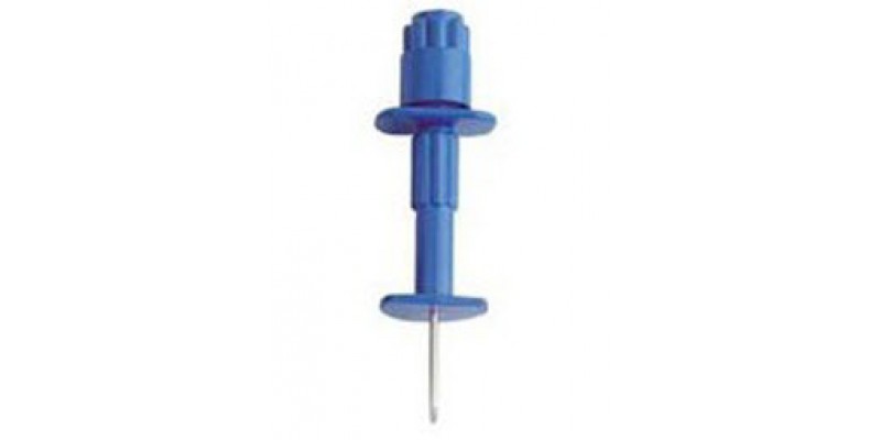
Equipment
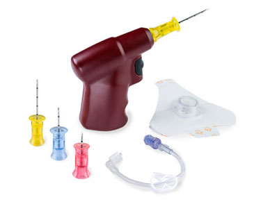
Several devices exist for IO insertion, including First Access for Shock and Trauma (FAST1), the EZ-IO, and the Bone Injection Gun (BIG). Manual devices include the Jamshidi needle and the Diekman modified needle. Clinicians should refer to the instruction manual from the manufacturer with the device for specific details on proper use. All have similar applications and techniques, except the sternal site, which requires the purchase of the FAST1 product to avoid penetration of the posterior cortex into the thoracic aorta.
Preparation
A standard sterile technique should be employed. The appropriate site is located, and if the patient is awake, although not mandatory, a local anesthetic can be injected into the site. The procedure is usually well-tolerated in the awake patient.
Technique
The needle is placed perpendicular to the bone with special attention in the pediatric population to avoid the epiphyseal plate. In pediatric placement, placement should be in the medial proximal tibia similar to the adult but 1 cm distal to the tibial tuberosity. Once the needle contracts the bone with the needle, a hard stop is felt, at least 5 mm of the needle should be visible above the skin to allow for penetration of the medullary space; if not, then choose a longer needle or a site with less soft tissue covering the bone. This scenario may occur with the obese patient, where the proximal tibial site is likely to be shallower.
Confirm placement of the IO needle by checking for the stability of needle in bone, aspiration of marrow, ability to flush with saline, and good IV flow rates. The inability to aspirate does not always indicate poor placement. If this occurs, continue with a saline flush and attempt aspiration again.
After needle insertion, flush with 5 to 10 cc normal saline for adults and 2 to 5 ml for infants and children. Patients may have severe pain when flushing with saline. The Injection of 2% intravenous lidocaine 20 to 40 mg or 0.5 mg/kg pediatric dose into the IO needle for relief of injection pain has been advocated, but with mixed results on pain resolution. Allow the Lidocaine 2 minutes to take effect before flushing.
Stabilization of the needle differs with each device used but is mandatory to avoid inadvertently dislodging or bending the IO needle.
The team should document the date and time of placement of the IO to ensure it will be in use for less than 24 hours. Following the obtaining of adequate IV access, the IO device should be removed and bandaged.

Complications
The inability to inject at the site indicates incomplete penetration of the needle through the cortex into the medullary space. This situation will be apparent due to the inability to flush with saline. Drilling deeper will resolve this problem.
Extravasation of fluid occurs secondary to drilling through the posterior cortex, placing an IO in a fracture site or in the bone that was previously accessed or at a recent orthopedic surgical site; this can cause compartment syndrome as well as the usual soft tissue complications similar to venous infiltration, depending on the product infused.
In the pediatric population, epiphyseal plate necrosis is avoidable by ensuring an IO insertion site is away from the epiphyseal plate.
Other complications include fracture, cellulitis, osteomyelitis, fat embolism, and the inability to remove a bent IO needle. A bent needle may require surgical removal.
Clinical Significance
IO access is fast and reliable with few complications and can be accomplished rather easily in the pre-hospital setting, with bedside training and by nursing staff and physicians. IV access is the preferred route, but nurses and physicians should not delay IO access in emergent situations if IV access is limited or will delay care.
Intraosseous aspiration of blood is usable for laboratory tests. The first aspirate does not need to be discarded. Studies vary on the accuracy of these results on hemodynamically stable and unstable animal sources. More comprehensive and standardized human studies in hemodynamically stable and unstable patients are needed to prove correlations with standard venous sampling – this is also true regarding the need to validate the values in point of care rapid testing. Following the obtaining of IV access, laboratory data should be repeated, ensuring accurate results.
Animal studies have shown expected values for blood gases, hematocrit, creatinine, sodium, and creatinine kinase in stable and hemorrhagic swine, but lactate and glucose values vary. Potassium values seem to be generally higher in IO samples compared with arterial and venous values in several studies. Blood typing is accurate even after blood transfusion through an IO device in a patient in shock, allowing type and crossing to be performed even after emergent transfusion.
