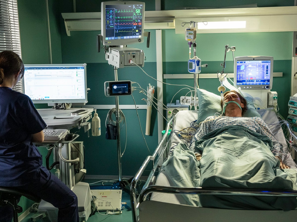
How Shock Patients Die: Acute Respiratory Distress Syndrome
Published .

Introduction
Acute respiratory distress syndrome (ARDS) is an acute, diffuse, inflammatory form of lung injury and life-threatening condition in seriously ill patients, characterized by poor oxygenation, pulmonary infiltrates, and acute onset. On a microscopic level, the disorder is associated with capillary endothelial injury and diffuse alveolar damage.
ARDS is an acute disorder that starts within seven days of the inciting event and is characterized by bilateral lung infiltrates and severe progressive hypoxemia in the absence of any evidence of cardiogenic pulmonary edema. ARDS is defined by the patient’s oxygen in arterial blood (PaO2) to the fraction of the oxygen in the inspired air (FiO2). These patients have a PaO2/FiO2 ratio of less than 300.
Once ARDS develops, patients usually have varying degrees of pulmonary artery vasoconstriction and may subsequently develop pulmonary hypertension. ARDS carries a high mortality, and few effective therapeutic modalities exist to combat this condition.
Etiology
ARDS has many risk factors. Besides pulmonary infection or aspiration, extra-pulmonary sources include sepsis, trauma, massive transfusion, drowning, drug overdose, fat embolism, inhalation of toxic fumes, and pancreatitis. These extra-thoracic illnesses and/or injuries trigger an inflammatory cascade culminating in pulmonary injury.
A lung injury prevention score helps identify low-risk patients, but a high score is less helpful.
Some risk factors for ARDS include:
- Advanced age
- Female gender
- Smoking
- Alcohol use
- Aortic vascular surgery
- Cardiovascular surgery
- Traumatic brain injury
- Pancreatitis
- Pulmonary contusion
- Infectious pneumonia
- Drugs (radiation, chemotherapeutic agents, amiodarone)
Epidemiology
Estimates of the incidence of ARDS in the United States range from 64.2 to 78.9 cases/100,000 person-years. Twenty-five percent of ARDS cases are initially classified as mild, and 75% as moderate or severe. However, a third of the mild cases progress to moderate or severe disease. Approximately 10 to 15% of patients admitted to the intensive care units and up to 23% of mechanically ventilated patients meet the criteria for ARDS.
Pathophysiology
ARDS represents a stereotypic response to various etiologies. It progresses through different phases, starting with alveolar-capillary damage, a proliferative phase characterized by improved lung function and healing, and a final fibrotic phase signaling the end of the acute disease process. The pulmonary epithelial and endothelial cellular damage is characterized by inflammation, apoptosis, necrosis, and increased alveolar-capillary permeability, which leads to the development of alveolar edema and proteinosis. Alveolar edema, in turn, reduces gas exchange, leading to hypoxemia. A hallmark of the pattern of injury seen in ARDS is that it is not uniform. Segments of the lung may be more severely affected, resulting in decreased regional lung compliance, which classically involves the bases more than the apices—this intrapulmonary differential in pathology results in a variant response to oxygenation strategies. While increased positive end-expiratory pressure (PEEP) may improve oxygen diffusion in affected alveoli, it may result in deleterious volutrauma and atelectrauma of adjacent unaffected alveoli. The injury results in three main outcomes:
- Impaired gas exchange
- Decreased lung compliance
- Pulmonary hypertension
Histopathology
The key histologic changes in ARDS reveal the presence of alveolar edema in diseased lung areas. The type I pneumocytes and vascular endothelium are injured, which results in the leaking of proteinaceous fluid and blood into the alveolar airspace. Other findings may include alveolar hemorrhage, pulmonary capillary congestion, interstitial edema, and hyaline membrane formation. None of these changes are specific to the disease.
History and Physical
The syndrome is characterized by dyspnea and hypoxemia, progressively worsening within 6 to 72 hours of the inciting event, frequently requiring mechanical ventilation and intensive care unit-level care. The history is directed at identifying the underlying cause that precipitated the disease. When interviewing patients that can communicate, they often start to complain of mild dyspnea initially, but within 12 to 24 hours, the respiratory distress escalates, becoming severe and requiring mechanical ventilation to prevent hypoxia. The etiology may be obvious in the case of pneumonia or sepsis. However, in other cases, questioning the patient or relatives on recent exposures may also be paramount in identifying the causative agent.
The physical examination will include findings associated with the respiratory system, such as tachypnea and increased breathing effort. Systemic signs may also be evident depending on the severity of the illness, such as central or peripheral cyanosis resulting from hypoxemia, tachycardia, and altered mental status. Despite 100% oxygen, patients have low oxygen saturation. Chest auscultation usually reveals rales, especially bibasilar, but can often be auscultated throughout the chest.
Differential Diagnosis
- Cardiogenic edema
- Exacerbation of interstitial lung disease
- Acute interstitial pneumonia
- Diffuse alveolar hemorrhage
- Acute eosinophilic lung disease
- Organizing pneumonia
- Bilateral pneumonia
- Pulmonary vasculitis
- Cryptogenic organizing pneumonia
- Disseminated malignancy
Prognosis
The prognosis for ARDS was abysmal until very recently. There are reports of 30 to 40% mortality up until the 1990s, but over the past 20 years, there has been a significant decrease in the mortality rate, even for severe ARDS. These accomplishments are secondary to a better understanding of and advancements in mechanical ventilation and earlier antibiotic administration and selection. The primary cause of death in patients with ARDS was sepsis or multiorgan failure. While mortality rates are now around 9 to 20%, it is much higher in older patients. ARDS has significant morbidity as these patients remain in the hospital for extended periods and have significant weight loss, poor muscle function, and functional impairment. Hypoxia from the inciting illness also leads to various cognitive changes that may persist for months after discharge. As measured by functional testing, there is an almost near-complete return of pulmonary capacity for many survivors. Nonetheless, many patients report feelings of dyspnea on exertion and decreased exercise tolerance. This ARDS sequela makes returning to a normal life challenging for these patients as they adjust to a new baseline.
Complications
- Barotrauma from high PEEP
- Prolonged mechanical ventilation -thus the need for tracheostomy
- Post-extubation laryngeal edema and subglottic stenosis
- Nosocomial infections
- Pneumonia
- Line sepsis
- Urinary tract infection
- Deep venous thrombosis
- Antibiotic resistance
- Muscle weakness
- Renal failure
- Post-traumatic stress disorder
