Shock
Published (updated: ).
Shock is a life-threatening manifestation of circulatory failure. Circulatory shock leads to cellular and tissue hypoxia resulting in cellular death and dysfunction of vital organs. Effects of shock are reversible in the early stages, and a delay in diagnosis and/or timely initiation of treatment can lead to irreversible changes, including multiorgan failure (MOF) and death.
Etiology
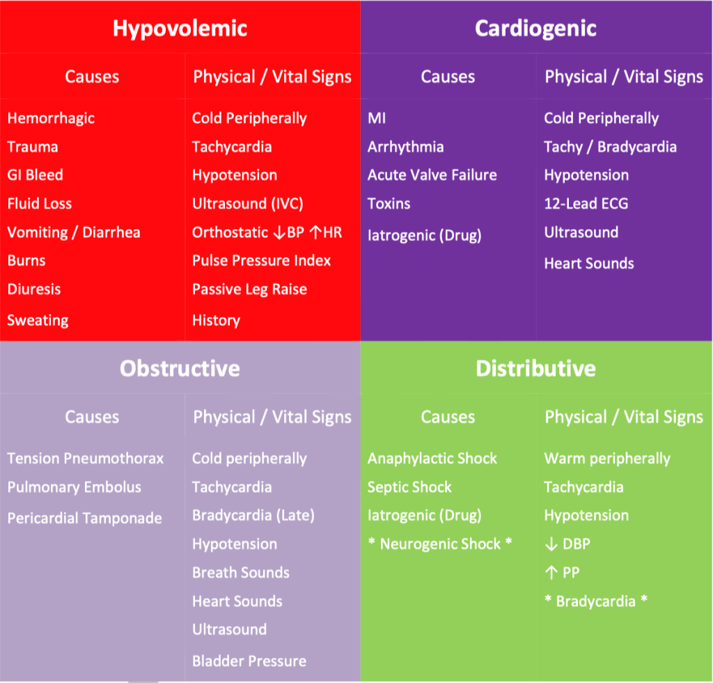
Shock is characterized by decreased oxygen delivery and/or increased oxygen consumption or inadequate oxygen utilization leading to cellular and tissue hypoxia. It is a life-threatening condition of circulatory failure and most commonly manifested as hypotension (systolic blood pressure less than 90 mm Hg or MAP less than 65 mmHg). Shock is the final manifestation of a complex list of etiologies and could be fatal without timely management. There are mainly four broad categories of shock: distributive, hypovolemic, cardiogenic, and obstructive. The wide range of etiologies can contribute to each of these categories and are manifested by the final outcome of shock. Undifferentiated shock means that the diagnosis of shock has been made; however, the underlying etiology has not been uncovered.
1. Distributive Shock
Characterized by peripheral vasodilatation.
Types of distributive shock include:
Septic Shock
Sepsis is defined as life-threatening organ dysfunction resulting from dysregulated host response to infection. Septic shock is a subset of sepsis with severe circulatory, cellular, and metabolic abnormalities resulting in tissue hypoperfusion manifested as hypotension which requires vasopressor therapy and elevated lactate levels.
The most common pathogens associated with sepsis and septic shock in the United States are gram-positive bacteria, including streptococcal pneumonia and Enterococcus.
Systemic Inflammatory Response Syndrome
Systemic inflammatory response syndrome (SIRS) is a clinical syndrome of the vigorous inflammatory response caused by either infectious or noninfectious causes. Infectious causes include pathogens such as gram-positive (most common) and gram-negative bacteria, fungi, viral infections (e.g., respiratory viruses), parasitic (e.g., malaria), rickettsial infections. Noninfectious causes of SIRS include but are not limited to pancreatitis, burns, fat embolism, air embolism, and amniotic fluid embolism
Anaphylactic Shock
Anaphylactic shock is a clinical syndrome of severe hypersensitivity reaction mediated by immunoglobulin E (Ig-E), resulting in cardiovascular collapse and respiratory distress due to bronchospasm. The immediate hypersensitivity reactions can occur within seconds to minutes after the presentation of the inciting antigen. Common allergens include drugs (e.g., antibiotics, NSAIDs), food, insect stings, and latex.
Neurogenic Shock
Neurogenic shock can occur in the setting of trauma to the spinal cord or the brain. The underlying mechanism is the disruption of the autonomic pathway resulting in decreased vascular resistance and changes in vagal tone.
2. Hypovolemic Shock
Hypovolemic shock is characterized by decreased intravascular volume and increased systemic venous assistance (compensatory the mechanism to maintain perfusion in the early stages of shock). In the later stages of shock due to progressive volume depletion, cardiac output also decreases and manifest as hypotension. Hypovolemic shock divides into two broad subtypes: hemorrhagic and non-hemorrhagic.
Common causes of hemorrhagic hypovolemic shock include
- Gastrointestinal bleed (both upper and lower gastrointestinal bleed (e.g., variceal bleed, portal hypertensive gastropathy bleed, peptic ulcer, diverticulosis) trauma
- Vascular etiologies (e.g., aortoenteric fistula, ruptured abdominal aortic aneurysm, tumor eroding into a major blood vessel)
- Spontaneous bleeding in the setting of anticoagulant use (in the setting of supratherapeutic INR from drug interactions)
Common causes of non-hemorrhagic hypovolemic shock include:
- GI losses – the setting of vomiting, diarrhea, NG suction, or drains.
- Renal losses – medication-induced diuresis, endocrine disorders such as hypoaldosteronism.
- Skin losses/insensible losses – burns, Stevens-Johnson syndrome, Toxic epidermal necrolysis, heatstroke, pyrexia.
- Third-space loss – in the setting of pancreatitis, cirrhosis, intestinal obstruction, trauma.
3. Cardiogenic Shock
Due to intracardiac causes leading to decreased cardiac output and systemic hypoperfusion. Different subtypes of etiologies contributing to cardiogenic shock include:
- Cardiomyopathies – include acute myocardial infarction affecting more than 40% of the left ventricle, acute myocardial infarction in the setting of multi-vessel coronary artery disease, right ventricular myocardial infarction, fulminant dilated cardiomyopathy, cardiac arrest (due to myocardial stunning), myocarditis.
- Arrhythmias – both tachy- and bradyarrhythmias
- Mechanical – severe aortic insufficiency, severe mitral insufficiency, rupture of papillary muscles, or chordae tendinae trauma rupture of ventricular free wall aneurysm.
4. Obstructive Shock
Mostly due to extracardiac causes leading to a decrease in the left ventricular cardiac output
- Pulmonary vascular – due to impaired blood flow from the right heart to the left heart. Examples include hemodynamically significant pulmonary embolism, severe pulmonary hypertension.
- Mechanical – impaired filling of right heart or due to decreased venous return to the right heart due to extrinsic compression. Examples include tension pneumothorax, pericardial tamponade, restrictive cardiomyopathy, constrictive pericarditis.
Epidemiology
Distributive shock is the most common type of shock, followed by hypovolemic and cardiogenic shock. Obstructive shock is relatively less common. The most common type of distributive shock is septic shock and has a mortality rate between 40 to 50%.
Pathophysiology
Hypoxia at the cellular level causes a series of physiologic and biochemical changes, resulting in acidosis and a decrease in regional blood flow, which further worsens the tissue hypoxia. In hypovolemic, obstructive, and cardiogenic shock, there is a decrease in cardiac output and decreased oxygen transport. In distributive shock, there is decreased peripheral vascular resistance and abnormal oxygen extraction. Excitement is a spectrum of physiologic changes, ranging from early stages, which are reversible to the final stages, which are irreversible with multiorgan failure and death. Generally, shock has the following three stages:
- Pre-shock or compensated shock – As the name suggests, this stage is characterized by compensatory mechanisms to counter the decrease in tissue perfusion, including tachycardia, peripheral vasoconstriction, and changes in systemic blood pressure
- Shock or decompensated shock – During this stage, most of the classic signs and symptoms of shock appear due to early organ dysfunction, resulting from the progression of the pre-shock stage as the compensatory mechanisms become insufficient.
- End-organ dysfunction of decompensated shock – This is the final stage, leading to irreversible organ dysfunction, multiorgan failure, and death
History and Physical
A focused history should be obtained from the patient (if feasible) and/or patient’s relatives. Also, a review of the patient’s outpatient medical records (information regarding risk factors, medications, and trend of baseline vital signs including blood pressure), as well as hospital medical records, could give valuable clues regarding the patient’s risk for shock and potential etiology. Clinical features and symptoms can vary according to the type and stage of shock. The most common clinical features/labs which are suggestive of shock include hypotension, tachycardia, tachypnea, obtundation or abnormal mental status, cold, clammy extremities, mottled skin, oliguria, metabolic acidosis, and hyperlactatemia. Also, features pertaining to the underlying cause of the shock can be present.
Patients with hypovolemic shock can have general features as mentioned above as well as evidence of orthostatic hypotension, pallor, flattened jugular venous pulsations, may have sequelae of chronic liver disease (in case of variceal bleeding).
Patients with septic shock may present with symptoms suggestive of the source of infection (example-skin manifestations of primary infection such as erysipelas, cellulitis, necrotizing soft-tissue infections), and cutaneous manifestations of infective endocarditis.
Patients with anaphylactic shock can have hypotension, flushing, urticaria, tachypnea, hoarseness of voice, oral and facial edema, hives, wheeze, inspiratory stridor, and history of exposure to common allergens such as medications or food items the patient is allergic to or insect stings.
Tension pneumothorax should be suspected in a patient with undifferentiated shock who has tachypnea, unilateral pleuritic chest pain, absent or diminished breath sounds, tracheal deviation to the normal side, distended neck veins and also has pertinent risk factors for tension pneumothorax such as recent trauma, mechanical ventilation, underlying cystic lung disease).
In a patient with undifferentiated shock, diagnostic clues to pericardial tamponade as the etiology include dyspnea, the Beck triad (elevated jugular venous pressure, muffled heart sounds, hypotension), pulses paradoxus, and known risk factors such as trauma, the recent history of pericardial effusion, and thoracic procedures.
Cardiogenic shock should be considered as the etiology if the patient with undifferentiated shock had chest pain suggestive of cardiac origin, narrow pulse pressure, elevated jugular venous pulsations or lung crackles, and significant arrhythmias on telemetry or EKG.
Evaluation
Resuscitation should not delay while investigating the etiology of undifferentiated shock. Physicians should have a high clinical suspicion for the presence of shock, and an attempt to stratify the severity of the shock should also take place to assess the need for emergent or early interventions. Evaluation of undifferentiated shock should begin with a thorough history and physical examination.
Besides telemetry monitoring, a 12-lead electrocardiogram should be obtained. ECGs might show evidence of acute coronary syndrome, arrhythmias, or provide diagnostic clues suggestive of pericardial effusion or pulmonary embolism.
Treatment / Management
The initial approach to management is the stabilization of the airway and breathing with oxygen and oral mechanical ventilation when needed. Peripheral IV or intraosseous infusion (IO) access should be obtained. Immediate treatment with intravenous (IV) fluid should be initiated, followed by vasopressor therapy, if needed, to maintain tissue perfusion. Depending on the underlying etiology of shock, specific therapies might also be needed.
Septic shock – initial aggressive fluid resuscitation with IV isotonic crystalloids 30 mL/kg within 3 hrs with additional fluid based on frequent reassessment, empiric antibiotic therapy within one hr.
Anaphylactic shock – aggressive IV fluid resuscitation with 2 liters of IV crystalloids. Stop the offending agent, intramuscular epinephrine, antihistamines, corticosteroids, nebulized albuterol.
Hypovolemic shock – obtain two large-bore IVs or central line. Place the patient in the Trendelenburg position. Aggressive IV fluid resuscitation with 2 liters of isotonic crystalloids. Packed red blood cell transfusion if ongoing bleed. Appropriate medical or interventional strategies to treat the underlying etiology.
Obstructive shock – the judicious use of IV crystalloids. Continue IV fluids but monitor very closely.
If acute massive pulmonary embolism -thrombolysis. Judicious use of IV fluids has a paradoxical worsening of hypotension; it may develop due to severe right ventricular dilatation and septal bowing compromising left ventricle filling.
If tension pneumothorax – needle thoracotomy followed by tube thoracotomy. If cardiac tamponade-pericardiocentesis, significant clinical improvement is possible, even with minimal fluid removal).
Cardiogenic shock – if unstable tachyarrhythmia or bradyarrhythmias, initiate ACLS protocol and cardioversion. Judicious use of IV fluids in the absence of pulmonary edema.
Pearls and Other Issues
- Shock is a clinical manifestation of circulatory failure and is associated with high morbidity and mortality.
- There are broadly four types of shock: distributive, cardiogenic, hypovolemic, and obstructive.
- An accurate diagnosis requires a good understanding of underlying pathophysiology, clinical, biochemical, and hemodynamic manifestations of the different types of shock.
- Serum lactate level is a useful risk stratification tool in managing undifferentiated shock.
- Timely diagnosis and initiation of appropriate therapy are of paramount importance as it can prevent progression to the reversible shock, multiorgan failure, and death.
- Treatment includes hemodynamic stabilization and correction of underlying etiology of shock.
