The 4 Chambered 2 Stage Pump
Published (updated: ).
Your heart is at the center of your circulatory system. This system is a network of blood vessels, such as arteries, veins, and capillaries, that carries blood to and from all areas of your body. Your blood carries the oxygen and nutrients that your organs need to work properly. Blood also carries carbon dioxide to your lungs so you can breathe it out. Inside your heart, valves keep blood flowing in the right direction.
Your heart’s electrical system controls the rate and rhythm of your heartbeat. A healthy heart supplies your body with the right amount of blood at the rate needed to work well. If disease or injury weakens your heart, your body’s organs will not receive enough blood to work normally. A problem with the electrical system — or the nervous or endocrine systems, which control your heart rate and blood pressure — can also make it harder for the heart to pump blood.
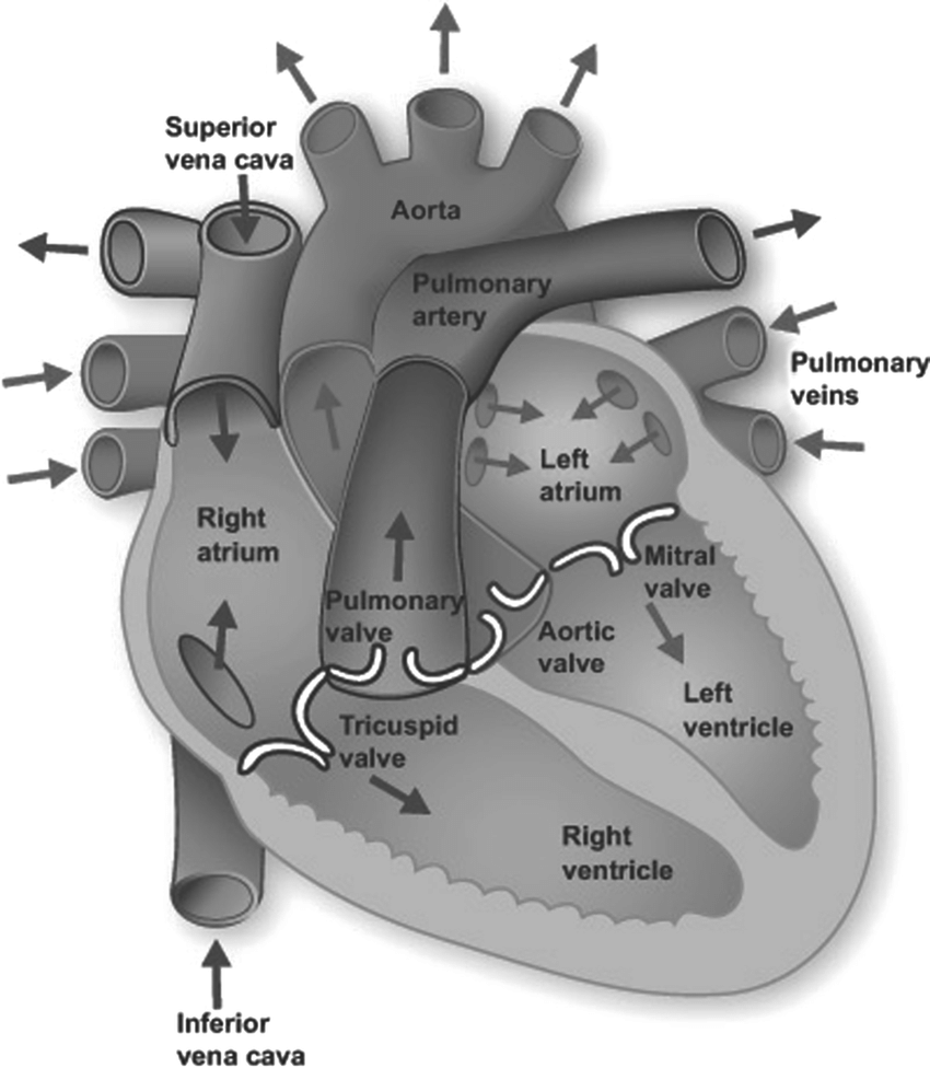
Your heart is in the center of your chest, near your lungs. It has four hollow chambers surrounded by muscle and other heart tissue. The chambers are separated by heart valves, which make sure that the blood keeps flowing in the right direction
Heart chambers
The two upper chambers of your heart are called atria, and the two lower chambers are called ventricles. Blood flows from the body and lungs to the atria and from the atria to the ventricles. The ventricles pump blood out of the heart to the lungs and other parts of the body. An internal wall of tissue divides the right and left sides of your heart. This wall is called the septum.
Heart tissue
The heart is made of three layers of tissue.
- Endocardium is the thin inner lining of the heart chambers and also forms the surface of the valves.
- Myocardium is the thick middle layer of muscle that allows your heart chambers to contract and relax to pump blood to your body.
- Pericardium is the sac that surrounds your heart. Made of thin layers of tissue, it holds the heart in place and protects it. A small amount of fluid between the layers helps reduce friction between the beating heart and surrounding tissues.
Some conditions can affect the heart’s tissue.
- Cardiomyopathy is when the heart muscle becomes enlarged, thick, or rigid. As cardiomyopathy worsens, the heart becomes weaker and is less able to pump blood through the body and maintain a normal electrical rhythm.
- Heart inflammation is inflammation in one or more of the layers of tissue in the heart, including the pericardium, myocardium, or endocardium. This can lead to serious complications, including heart failure, cardiogenic shock, or irregular heart rhythm.
Acute Inflammation
Tissue damage due to trauma, microbial invasion, or noxious compounds can induce acute inflammation. Inflammation is part of the body’s defense mechanism. It is the process by which the immune system recognizes and removes harmful and foreign stimuli and begins the healing process. Inflammation can be either acute or chronic. It starts rapidly, becomes severe in a short time and symptoms may last for a few days for example cellulitis or acute pneumonia. Subacute inflammation is the period between acute and chronic inflammation and may last 2 to 6 weeks.
Chronic Inflammation
Chronic inflammation is also referred to as slow, long-term inflammation lasting for prolonged periods of several months to years. Generally, the extent and effects of chronic inflammation vary with the cause of the injury and the ability of the body to repair and overcome the damage.
- Congenital heart disease is when the heart does not develop in the typical way. A congenital heart defect can happen at any point during development of an unborn baby, or embryo, inside the pregnant mother.
Arteries and veins link your heart to the rest of the circulatory system. Veins bring blood to your heart. Arteries take blood away from your heart. Your heart valves help control the direction the blood flows.
Heart valves
Heart valves control the flow of blood so that it moves in the right direction. The valves prevent blood from flowing backward.
The heart has four valves.
- The tricuspid valve separates the right atrium and right ventricle.
- The mitral valve separates the left atrium and left ventricle.
- The pulmonary valve separates the right ventricle and the pulmonary artery.
- The aortic valve separates the left ventricle and aorta.
The valves open and shut in time with the pumping action of your heart’s chambers. The opening and closing involve a set of flaps called cusps or leaflets. The cusps open to allow blood to flow out of a chamber and close to allow the chamber to refill with blood. Heart valve diseases can cause backflow or slow the flow of blood through the heart.
Adding oxygen to blood
Oxygen-poor blood from the body enters your heart through two large veins called the superior and inferior vena cava. The blood enters the heart’s right atrium and is pumped to your right ventricle, which in turn pumps the blood to your lungs.
The pulmonary artery then carries the oxygen-poor blood from your heart to the lungs. Your lungs add oxygen to your blood. The oxygen-rich blood returns to your heart through the pulmonary veins.
The oxygen-rich blood from the lungs then enters the left atrium and is pumped to the left ventricle. The left ventricle generates the high pressure needed to pump the blood to your whole body through your blood vessels.
When blood leaves the heart to go to the rest of the body, it travels through a large artery called the aorta. A balloon-like bulge, called an aortic aneurysm, can sometimes occur in the aorta.
Supplying oxygen to the heart’s muscle
Like other muscles in the body, your heart needs blood to get oxygen and nutrients. Your coronary arteries supply blood to your heart. These arteries branch off from the aorta so that oxygen-rich blood is delivered to your heart as well as the rest of your body.
- The left coronary artery delivers blood to the left side of your heart, including your left atrium and ventricle and the septum between the ventricles.
- The circumflex artery branches off from the left coronary artery to supply blood to part of the left ventricle.
- The left anterior descending artery also branches from the left coronary artery and provides blood to parts of both the right and left ventricles.
- The right coronary artery provides blood to the right atrium and parts of both ventricles.
- The marginal arteries branch from the right coronary artery and provide blood to the surface of the right atrium.
- The posterior descending artery also branches from the right coronary artery and provides blood to the bottom of both ventricles.
Some conditions can affect normal blood flow through these heart arteries. Examples include:
- Angina
- Heart attack
- Coronary heart disease
The coronary veins return oxygen-low blood from the heart’s muscles back to the right atrium so it can be pumped to the lungs. They include:
- The anterior cardiac veins
- The great cardiac vein
- The middle cardiac vein
- The small cardiac vein
Your heartbeat is the contraction of your heart to pump blood to your lungs and the rest of your body. Your heart’s electrical system determines how fast your heart beats.
Your heartbeat
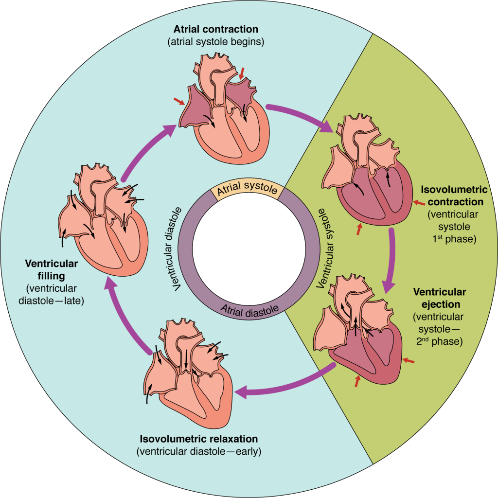
The contraction of the atria and ventricles makes a heartbeat. When your heart beats, it makes a “lub-DUB” sound. You may have heard this if you listened with a stethoscope or with your ear on someone’s chest.
- After your atria pump blood into the ventricles, the valves between the atria and ventricles close to prevent backflow. The “lub” is the sound of these valves closing.
- After your ventricles contract to pump blood away from the heart, the aortic and pulmonary valves close and make the “dub” sound.
What is a normal pulse?
Your pulse is the rate your heart beats. It is also called your heart rate. To find your pulse without a heartrate monitor or watch, gently place your index and middle fingers on the artery located on the inner wrist of either arm, below your thumb. You should feel a pulsing or tapping against your fingers.
Watch the second hand or set the timer on your stopwatch or phone and count the number of beats you feel in 30 seconds. Double that number to find out your heart rate or pulse for 1 minute.
- At rest, a heart rate of 60 to 100 beats per minute is normal.
- When you exercise, your heart beats faster, and your heart rate speeds up to get more oxygen to your muscles.
Signals from your body’s nervous system and hormones from your endocrine system control how fast and hard your heart beats. These signals and hormones allow you to adapt to changes in the amount of oxygen and nutrients your body needs.
Electrical activity
Electrical signals cause muscles to contract. Your heart has a special electrical system called the cardiac conduction system. This system controls the rate and rhythm of the heartbeat.
With each heartbeat, an electrical signal travels from the top of the heart to the bottom. As the signal travels, it causes the heart to contract and pump blood. The heartbeat process includes the following steps.
- The signal begins in a group of cells, called pacemaker cells, located in the sinoatrial (SA) node in the right atrium.
- The electrical signal travels through the atria, causing them to pump blood into the ventricles.
- The electrical signal then moves down to a group of pacemaker cells called the atrioventricular (AV) node, located between the atria and the ventricles. Here the signal slows down slightly, allowing the ventricles time to finish filling with blood.
- The AV node fires another signal that travels along the walls of your ventricles, causing them to contract and pump blood out of your heart.
- The ventricles relax, and the heartbeat process starts all over again in the SA node.
Some conditions affect the heart’s electrical system. Examples are included below.
- Arrhythmia is an irregular heart rhythm. Atrial fibrillation is one of the most common types of arrhythmia.
- Conduction disorders can happen when electrical signals either do not generate properly, do not travel properly through the heart, or both.
Blood pressure
Your blood pressure is the force of the blood pushing against the walls of your arteries as the heart pumps blood. It is made up of two numbers: systolic and diastolic.
- Systolic pressure is the pressure when the ventricles pump blood out of the heart. The pressure on your arteries is highest during this time.
- Diastolic pressure is the pressure between beats, when the heart is filling with blood. The pressure on your arteries is lowest during this time.
For most adults, healthy blood pressure is usually less than 120 over 80, which is written as your systolic pressure number over your diastolic pressure number.
High blood pressure is what happens when blood flows through blood vessels at higher-than-normal pressures.
