Airway Burns
Published .
Introduction
Inhalation injury is a broad term that includes pulmonary exposure to a wide range of chemicals in various forms including smoke, gases, vapors, or fumes. Inhalation injury from smoke exposure is commonly seen in patients exposed to fires. Smoke inhalation is one of the most commonly encountered inhalation injuries and is the primary focus of this information. Information relating to external thermal burns and caustic ingestions will not be discussed.
Etiology
Smoke inhalation injuries occur when a patient’s respiratory system is exposed to direct heat from fire as well as toxic chemicals that are formed from the decomposition of materials during combustion. The composition of smoke varies with each fire depending upon the materials being burned, the amount of oxygen available to the fire, and the nature of the fire. High-oxygen and high-temperature fires often do not produce large amounts of smoke. Low oxygen fires are often lower temperature fires, and these lower temperatures often give rise to more toxic chemicals, such as carbon monoxide. Other common toxic compounds created in smoke are ammonia, carbon dioxide, hydrogen cyanide, aldehydes, sulfur dioxides, and nitrogen dioxide. These different elements give rise to a combination of gases, airborne solids, and liquid vapors that mix with the ambient air to create smoke. Inhalation of these components, when exposed to smoke, causes both upper and lower airway injury.
Epidemiology
According to FEMA records, in 2015, there were 380,940 residential fires, resulting in 2565 deaths, and 11,475 fire-related injuries in the United States. The number of deaths has not decreased since 2006 and has trended 2% upward despite a decrease of 9% in fire injuries during this same period. The leading cause of death from fire injuries remains respiratory failure, and smoke inhalation injuries affect one-third of all burn injury victims.
Pathophysiology
Inhalation injury affects the respiratory system through damage to the airways (including nasal passages, posterior oropharynx, larynx, trachea, bronchi) or parenchymal damage (alveoli). The location where damage occurs is complex. Thermal injury often affects only to the level of the larynx. Chemical toxin/irritants may cause damage to just the airways, just the alveoli, or both. Specifically, water solubility for gases or vapors, and the physical characteristics of the particulates for fumes and aerosols are important for determining the location of the injury. More water-soluble chemicals will often damage the moist mucosa of the upper airway without causing alveolar damage. Examples of highly water-soluble chemicals include ammonia and sulfur dioxide. Chemical toxins that have low water solubility may reach the lung parenchyma without damage to the airways.
Damage to airway tissue causes increased mucus production, edema, denudation of epithelium, and mucosal ulceration and hemorrhage. Obstruction of airflow is often the effect caused by tissue edema narrowing the passageways and mucus/blood/fluid impeding airflow. Pseudomembranes may also form in the trachea or bronchi causing bronchiolitis obliterans and organizing pneumonia. Damage to the lung parenchyma causes both epithelial and endothelial damage resulting in pulmonary edema and possibly acute respiratory distress syndrome (ARDS) due to widespread alveolar-capillary leak. A direct thermal injury is rare past the vocal cords because even superheated air is quickly cooled by the nasopharynx and oropharynx prior to causing lower respiratory tract injury. Most lower respiratory tract injury is from smoke particles and the chemicals that they carry.
Histopathology
The histopathology of inhalation parenchymal injury is diffuse alveolar damage (DAD): diffuse edema with epithelial necrosis and cell sloughing. After this initial phase, hyaline membrane formation occurs. Finally, DAD may organize, leading to the proliferation of type II pneumocytes with resorption of the hyaline membranes and exudates, and fibroblast proliferation. The long-term effects of DAD include both complete recovery and permanent interstitial fibrosis. Many inhaled chemical toxins cause DAD.
History and Physical
A high index of suspicion is important for all clinicians to have when evaluating patients for inhalation injury. It is important to elucidate whether the exposure was to smoke, flames, and/or possible chemicals (both industrial and household). Duration of exposure, the location of exposure (such as if it was in an enclosed space), and any loss of consciousness are all important as well. Patients may be unconscious at presentation and interviewing first responders/rescuers may be required. Duration of exposure is often greater for pediatric and elderly patients as often they will have longer exposure due to disorientation or mobility issues. Children also often hide from smoke or fires thus increasing time exposed. Pediatric patients also have increased minute ventilation with a higher respiratory rate when compared to adults, thus, increasing the amount of exposure.
History taking should be complete and thorough. Burn patients may have extensive external injuries, but smoke inhalation may affect those with no outward signs of burns. Patients suffering from smoke inhalation may have symptoms of burning sensation in the nose or throat (which is often caused by an irritant chemical toxin), a cough with increased sputum production, stridor, and dyspnea with rhonchi or wheezing. Symptoms of odynophagia after smoke exposure should also raise suspicion for a possible inhalation injury. Patients may have systemic symptoms like a headache, delirium, hallucinations, and may even be comatose. Many different etiologies may cause changes in mental status including hypoxia, hypercarbia, or asphyxiant exposure (carbon monoxide, hydrogen cyanide). In cases where history is limited, some physical examination findings may cue the examiner into possible inhalation injury.
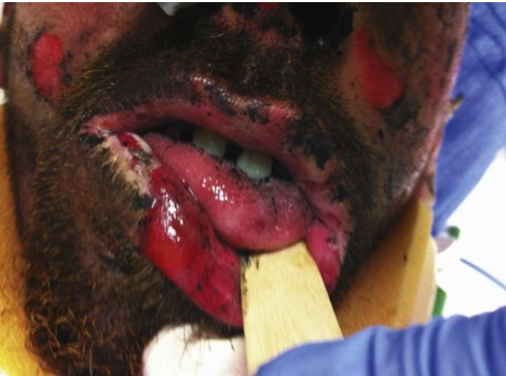
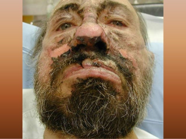
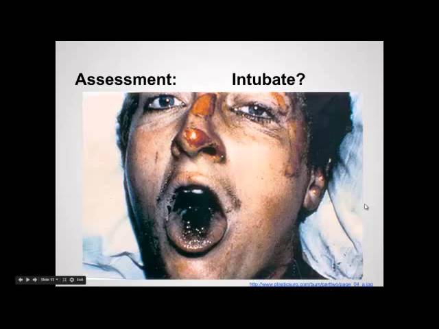
Physical examination should include looking for facial burns, such as loss of facial and intranasal hair as well as carbonaceous material or soot in the mouth or sputum. There may be accessory muscle usage, tachypnea, cyanosis, stridor, and rhonchi/rales/wheezing. Findings of stridor or upper airway turbulence/noise are often a sign of impending airway compromise, and prompt intubation should be strongly considered.
A delay in the onset of symptoms is not uncommon, and clinicians should educate patients on the possibility of delayed symptom onset post-exposure. The delayed symptoms occur in the lower respiratory airways as it is caused by chemical toxin exposure, which may bypass the upper airways. For example, nitric oxide is often produced by the burning of certain fabrics or cellulose. It is highly water-insoluble, and damage onset has been seen up to 72 hours post-exposure.
Evaluation
At the hospital, a typical work up of smoke inhalation injury may include the following:
- Chest imaging: Serial chest radiographs (often negative early in smoke inhalation injury), computed tomography (CT) chest
- Complete blood count (CBC), complete metabolic panel (CMP), lactate
- Pulse oximetry (may be falsely elevated with carbon monoxide exposure)
- Arterial blood gas (ABG)
- Carboxyhemoglobin level
- Cyanide level (often not readily available therefore limited use in the acute setting)
- Pulmonary function testing: The flow-volume loop is a very sensitive noninvasive test
- Bronchoscopy and direct laryngoscopy
Treatment / Management
Limiting exposure and removing the patient from the exposure area, such as in a house fire or occupational exposure, and maintaining a secure airway are paramount. Airway protection should include considering early and preemptive intubation for patients with inhalation injury. Airway edema may occur suddenly as edema worsens, and often, the upper airways develop injury and obstruction earliest, prior to the parenchymal injury. Treatment as a whole is largely supportive with specific therapies below:
- Maintain secure airway: Intubation, tracheostomy if necessary
- Obstruction often results in edema, hemorrhage, and mucosal sloughing, and aggressive pulmonary hygiene can help to manage these secretions.
- Obstruction may also occur due to airway reactivity, for which bronchodilators should be used. Beta-2-adrenergic agonists, including albuterol have been shown in animal models to improve pulmonary functioning in smoke inhalation injury.
- Usage of steroids, in either inhaled or intravenous form, has not been proven beneficial in clinical studies.
Specific treatments may vary depending on other factors, as often inhalation injury may present with systemic toxicities, for example, carbon monoxide (CO) and cyanide poisoning in smoke inhalation injury.
- Carbon monoxide treatment includes high oxygen therapy. Hyperbaric oxygen treatment (HBO) has been shown to increase the clearance rate of CO from blood, but its limited availability restricts its usage. Most health care centers instead use 100% FiO2 oxygen therapy for treatment.
- Hydrogen cyanide poisoning is difficult to determine in many cases. Testing results are not readily available. One antidote, hydroxocobalamin, may be given in patients with high suspicion of cyanide poisoning. It is nontoxic and renally excreted. Side effects do include redding of the skin and urine.
Accurate treatment of possible toxic inhalation injuries is accomplished by identifying the possible compounds inhaled, the duration and relative concentration of exposure, and the water solubility of any toxic inhaled agent.
Prognosis
Patients with smoke inhalation injury are at high risk for complications. The majority of fatal burn cases are caused by respiratory failure, either from direct injury or complications such as pneumonia. Severe injuries often will lead to long-term complications such as bronchiectasis, bronchiolitis obliterans, and the need for artificial airways; however, many patients will have no long-term sequelae from a single episode of smoke inhalation injury.
Complications
Most cases of smoke inhalation injury will be mild to moderate in severity. Severity generally correlates with exposure time. More serious injuries occur with longer and more intense exposure. Mild to moderate injury is largely self-limited with patients having no complications. Patient symptoms will often resolve within 2 to 3 days.
Short-term complications are seen in more severe injuries within 4 to 5 days, and the most common issue is pneumonia. Acute respiratory distress syndrome and pulmonary edema are also seen in the short term. These patients will often demonstrate changes in pulmonary function testing and may require ventilatory support. Complication rates are higher in people with a history of underlying lung disease as well, such as COPD and asthma.
Long-term complications from smoke inhalation injury are much less common. They include subglottic stenosis, bronchiectasis, and bronchiolitis obliterans. Patients who have been exposed to carbon monoxide are also known to have long-term neurological complications. Severe brain damage may occur with carbon monoxide poisoning but is uncommon. More commonly patients will describe persistent or delayed neurological symptoms after carbon monoxide poisoning. These symptoms are often subjective but will include depressed mood, poor concentration, and issues with short-term memory. Neurological sequelae after carbon monoxide exposure seem to be more common in patients who had a loss of consciousness. Symptoms often develop 1 to 3 weeks after the poisoning. Hyperbaric oxygen therapy is being investigated as a possible therapy for these neurological sequelae, but more research is needed in this area.
Deterrence and Patient Education
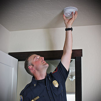
Prevention of smoke inhalation is achieved through means of fire prevention. Properly functioning fire and smoke alarms are the best means of preventing inhalation injury and fire injuries in general. Smoke detectors are prevalent in the United States, 96% of US households reportedly use smoke detectors; however, over 5 million homes still do not have smoke alarms. In 38% of home fire deaths, no smoke alarms were present, and in 21% of home fire deaths, smoke alarms did not sound, often because they were intentionally disconnected or had missing batteries.
Pearls and Other Issues
- Smoke inhalation injury occurs from either direct thermal injury, inhalation of irritants that damage respiratory tissues, or inhalation of chemicals with systemic effects/ asphyxiants.
- The majority of burn victim deaths are from respiratory failure.
- Smoke inhalation injury greatly increases mortality in burn patients.
- Longer exposure time is associated with more severe injury.
- Treatment is largely supportive, and a low threshold for intubation is important for patient survival.
- Hyperbaric oxygen therapy has shown some promise for inhalation injury, including carbon monoxide poisoning, but is not standard of care due and has limited availability.
- Primary prevention is the best deterrence of inhalation injury.
