Protective Layers Of The Brain
Published (updated: ).
The central nervous system (CNS) is crucial to the operation of the body and any compromise of function in the brain and spinal cord can lead to severe difficulties. The brain is protected by multiple structures. First, the bones of the skull enclose and house the brain. Underneath the skeletal structures, the brain is protected by membranes made by connective tissue, called meninges, that surround, support, stabilize and partition the nervous tissue. In addition, the brain has a privileged blood supply, as suggested by the blood-brain barrier. The function of the tissue is crucial to the survival of the organism, so the contents of the blood cannot simply pass into the central nervous tissue. To protect this region from the toxins and pathogens that may be traveling through the blood stream, there is strict control over what can move out of the general systems and into the brain. Because of this privilege, the brain needs specialized structures for the maintenance of circulation. This begins with a unique arrangement of blood vessels carrying fresh blood into the brain and venous sinuses carrying deoxygenated blood out of the brain. Beyond the supply of blood, the brain filters the blood into cerebrospinal fluid (CSF), which is then circulated through the cavities of the brain, such as the subarachnoid space and the ventricles.
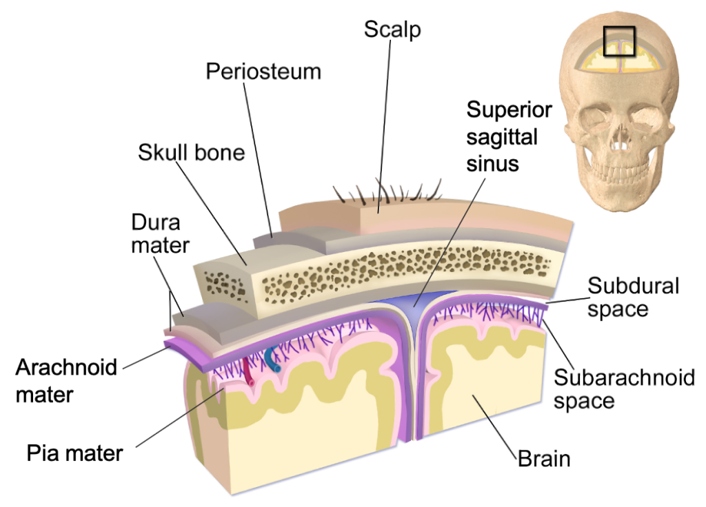
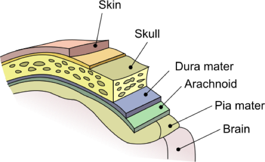
The outer surface of the CNS is covered by a series of membranes composed of connective tissue called the meninges, which protect, stabilize and partition the brain. From superficial to deep, the meningeal layers are the dura mater, arachnoid mater and pia mater. The dura mater is a thick fibrous layer and a strong protective sheath over the entire brain. It is anchored to the inner surface of the cranium and vertebral cavity. The arachnoid mater is a membrane of thin fibrous tissue that forms a loose sac around the brain. Beneath the arachnoid mater, there is a space called the subarachnoid space where a thin, filamentous mesh form the arachnoid trabeculae, that looking like a spider web give this layer its name. Directly adjacent to the surface of the brain is the pia mater, a thin fibrous membrane that follows the superficial convolutions of the brain and fits into other grooves and indentations.
Like a thick cap covering the brain, the dura mater is a tough outer covering. The name comes from the Latin for “tough mother” to represent its physically protective role. It encloses the entire CNS and the major blood vessels that enter the cranium and vertebral cavity. It is directly attached to the inner surface of the bones of the cranium and to the very end of the vertebral cavity. The dura mater of the brain is made by two layers, the periosteal layer which is more superficial and attached to the skull, and the meningeal layer, which lies deep to the first layer. These two layers are usually fused together. However, in some regions of the brain they separate to form large space filled with venous blood called dural sinuses. The dura mater of the spinal cord is composed of only one layer. The region between the bones and the dura mater is called epidural space. In the cranium, the epidural space contains arteries and veins that supply and drain blood from the brain. In normal conditions, the cranial epidural space is a potential space and not a real space. However, trauma can cause the leakage of fluid (hematomas) which accumulates in the epidural space enlarging it and transforming it into a real space. In the vertebral column, the epidural space is a real space that contains areolar and adipose connective tissue for an extra layer of protection, as well as blood vessels. The spinal epidural space at the lumbar level is where epidural injections are administered. Deep to the dura mater, there is another potential space called the subdural space. As the epidural space, this space can be filled with fluid causing a subdural hematoma.
The meningeal layer of the dura mater extends into the cranial cavity at four locations. These flat partitions are called cranial dural septa and separate specific parts of the brain. The falx cerebri goes through the midline separation of the brain while the falx cerebelli separates the two cerebellar hemispheres. The tentorium cerebelli separates the cerebellum from the superior part of the brain (cerebrum), forming a shelf-like tent. Another partition called diaphragma sellae surrounds the pituitary gland.
Deep to the subdural space lies the middle layer of the meninges called the arachnoid mater. This layer is named for the spider-web–like projections called arachnoid trabeculae between this layer and the pia mater. The arachnoid defines a sac-like enclosure around the CNS. The trabeculae are found in the subarachnoid space, which is filled with circulating CSF, making it a real space. The arachnoid emerges into the dural sinuses as the arachnoid granulations, where the CSF is filtered back into the blood for drainage from the nervous system.
The outer surface of the CNS is covered in the thin fibrous membrane of the pia mater. It is thought to have a continuous layer of cells providing a fluid-impermeable membrane. The name pia mater comes from the Latin for “tender mother,” suggesting the thin membrane is a gentle covering for the brain. The pia extends into every convolution of the CNS. Blood vessels that are nourishing the central nervous tissue are between the pia mater and the nervous tissue.
