Anatomy Of The Brain
Published (updated: ).
The brain controls many important body functions, such as emotions, vision, thought, speech, and movement. The spinal cord connects the brain to nerves in most parts of the body. This allows the brain to send messages throughout the body. The network of the brain and spinal cord is called the central nervous system (CNS).
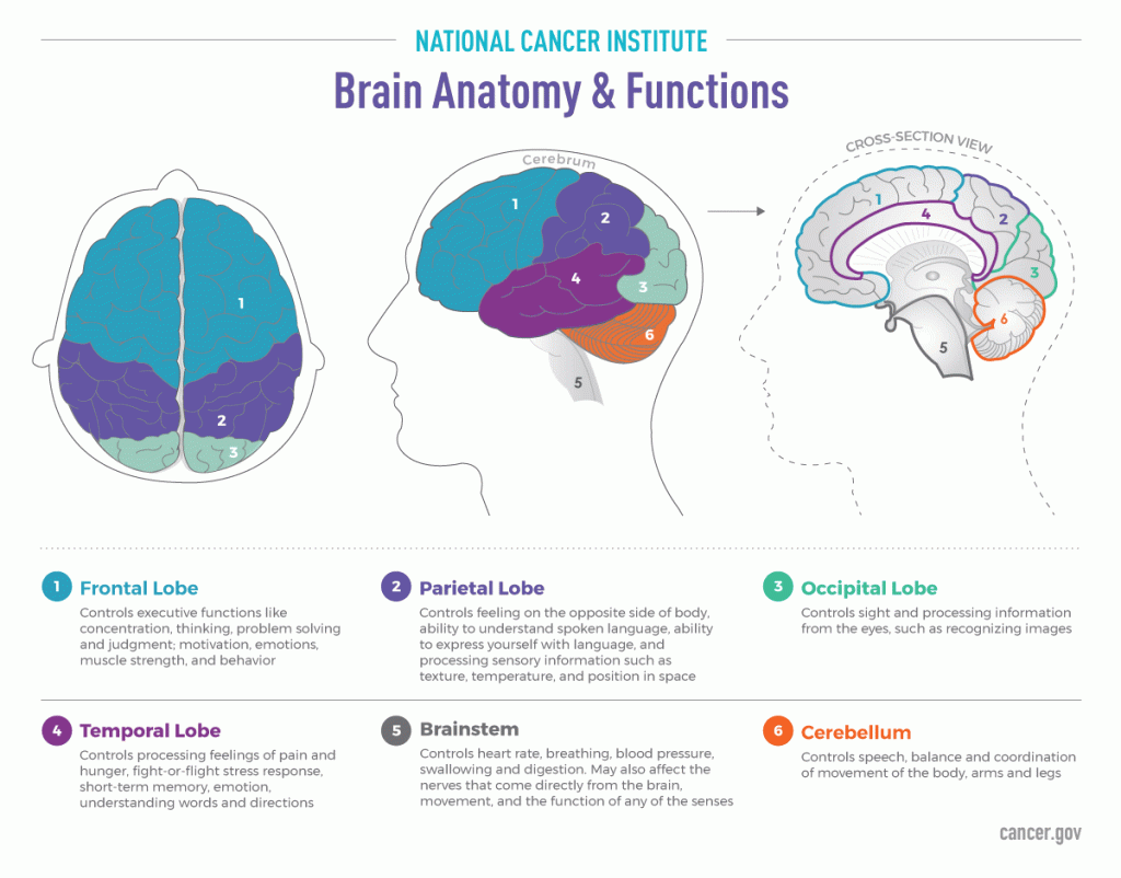
Brain Anatomy and Functions
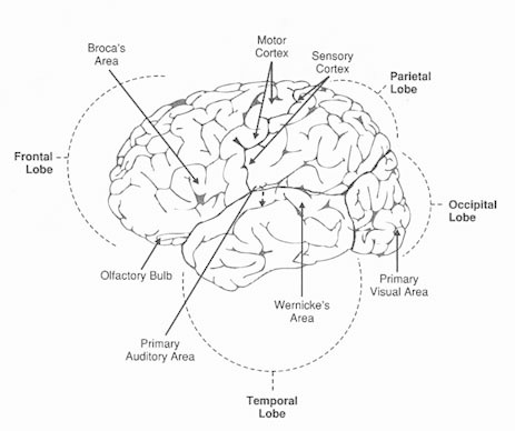
The brain is divided down the middle from front to back into two halves called the cerebral hemispheres. Each hemisphere is divided into four lobes: frontal, parietal, occipital, and temporal.
The preponderance of the cerebral cortex (which, with its supporting structures, makes up approximately 80 percent of the brain’s total volume) is actually a recent development in the course of evolution. The cortex contains the physical structures responsible for most of what we call ”brainwork”: cognition, mental imagery, the highly sophisticated processing of visual information, and the ability to produce and understand language. But underneath this layer reside many other specialized structures that are essential for movement, consciousness, sexuality, the action of our five senses, and more—all equally valuable to human existence. Indeed, in strictly biological terms, these structures can claim priority over the cerebral cortex. In the growth of the individual embryo, as well as in evolutionary history, the brain develops roughly from the base of the skull up and outward. The human brain actually has its beginnings, in the four-week-old embryo, as a simple series of bulges at one end of the neural tube.
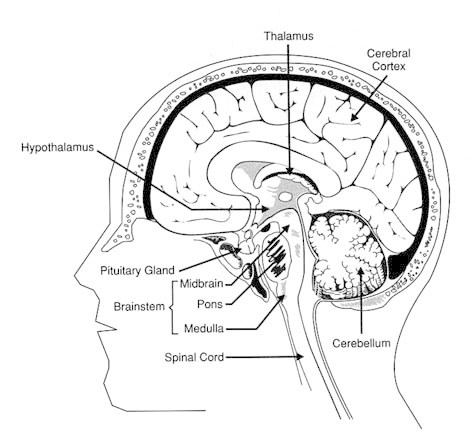
Ventricles
The bulges in the neural tube of the embryo develop into the hindbrain, midbrain, and forebrain—divisions common to all vertebrates, from sharks to squirrels to humans. The original hollow structure is commemorated in the form of the ventricles, which are cavities containing cerebrospinal fluid. During the course of development, the three bulges become four ventricles. In the hindbrain is the fourth ventricle, continuous with the central canal of the spinal cord. A cavity in the forebrain becomes the third ventricle, which leads further forward into the two lateral ventricles, one in each cerebral hemisphere.
Brainstem
The hindbrain contains several structures that regulate autonomic functions, which are essential to survival and not under our conscious control. The brainstem, at the top of the spinal cord, controls breathing, the beating of the heart, and the diameter of blood vessels. This region is also an important junction for the control of deliberate movement. Through the medulla, at the lower end of the brainstem, pass all the nerves running between the spinal cord and the brain; in the pyramids of the medulla, many of these nerve tracts for motor signals cross over from one side of the body to the other. Thus, the left brain controls movement of the right side of the body, and the right brain controls movement of the left side.
In addition to being the major site of crossover for nerve tracts running to and from the brain, the medulla is the seat of several pairs of nerves for organs of the chest and abdomen, for movements of the shoulder and head, for swallowing, salivation, and taste, and for hearing and equilibrium.
At the top of the brainstem is the pons—literally, a bridge—between the lower brainstem and the midbrain. Nerve impulses traversing the pons pass on to the cerebellum (or “little brain”), which is concerned primarily with the coordination of complex muscular movement. In addition, nerve fibers running through the pons relay sensations of touch from the spinal cord to the upper brain centers.
Many nerves for the face and head have their origin in the pons, and these nerves regulate some movements of the eyeball, facial expression, salivation, and taste. Together with nerves of the medulla, nerves from the pons also control breathing and the body’s sense of equilibrium.
What had been the middle bulge in the neural tube develops into the midbrain, which functions mainly as a relay center for sensory and motor nerve impulses between the pons and spinal cord and the thalamus and cerebral cortex. Nerves in the midbrain also control some movements of the eyeball, pupil, and lens and reflexes of the eyes, head, and trunk.
The “Little Brain” At The Back Of The Head
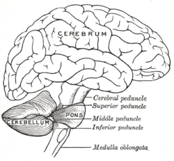
While autonomic and endocrine functions are being maintained by structures deep inside the brain, another specialized area is sorting and processing the signals required to maintain balance and posture and to carry out coordinated movement. The cerebellum (the term in Latin means “little brain”) is actually a derived form of the hindbrain—as suggested by its position at the back of the head, partly tucked under the cerebral hemispheres. In humans, with our almost unlimited repertoire of movement, the cerebellum is accordingly large; in fact, it is the second-largest portion of the brain, exceeded only by the cerebral cortex. Its great surface area is accommodated within the skull by elaborate folding, which gives it an irregular, pleated look. In relative terms, the cerebellum is actually largest in the brain of birds, where it is responsible for the constant streams of information between brain and body that are required for flight.
In humans, the cerebellum relays impulses for movement from the motor area of the cerebral cortex to the spinal cord; from there, they pass to their designated muscle groups. At the same time, the cerebellum receives impulses from the muscles and joints that are being activated and in some sense compares them with the instructions issued from the motor cortex, so that adjustments can be made (this time by way of the thalamus). The cerebellum thus is neither the sole initiator of movement nor a simple link in the chain of nerve impulses, but a site for the rerouting and in some cases refining of instructions for movement. There is evidence, too, that the cerebellum can store a sequence of instructions for frequently performed movements and for skilled repetitive movements—those that we think of as learned “by rote.”
The right and left hemispheres of the cerebellum each connect with the nerve tracts from the spinal cord on the same side of the body, and with the opposite cerebral hemisphere. For example, nerve impulses concerned with movement of the left arm originate in the right cerebral hemisphere, and information about the orientation, speed, and force of the movement is fed back to the right cerebral hemisphere, through the left half of the cerebellum. The nerves responsible for movement at the ends of the arms and legs tend to have their origin near the outer edges of the cerebellum. By contrast, nerves that have their origin near the center of the cerebellum serve to monitor the body’s overall orientation in space and to maintain upright posture, in response to information about balance that is transmitted by nerve impulses from the inner ear, among other sources.
Reticular Network
Some nerve fibers from the cerebellum also contribute to the reticular formation, a widespread network of neurons (“reticular” is derived from the Latin word for “net”). This formation and some neurons in the thalamus, together with others from various sensory systems of the brain, make up the reticular activating system—the means by which we maintain consciousness. The reticular activating system also comes into play when we deliberately focus our attention, “tuning out” distractions to some degree. At the midline of the brainstem are the raphe nuclei, whose axons extend down into the spinal cord and up to the cerebral cortex—a reach that makes it possible for many areas of the nervous system to be contacted simultaneously. The reticular formation plays a role in movement, particularly those forms of movement that do not call for conscious attention: it is also involved in transmitting or inhibiting sensations of pain, temperature, and touch. Less tangibly, the reticular activating system appears to work as a filter for the countless stimuli that can act on the nervous system both from within and from outside the body. It is this filtering of signals that allows a passenger on an airplane, for example, to doze off undisturbed by sounds of nearby conversation and steady jet engines, but to awake and become alert when the pitch of the engines changes and the plane tilts into its descent.
The “Emotional Brain”
The limbic system (from the Latin limbus, for “hem” or “border”) is another assembly of linked structures that form a loose circuit throughout the brain. This system is a fairly old part of the brain and one that humans share with many other vertebrates; in reptiles, it is known as the rhinencephalon, or “smell-brain,” because it reacts primarily to signals of odor. In humans, of course, the stimuli that can affect the emotional brain are just about limitless in their variety.
The limbic system is responsible for most of the basic drives and emotions and the associated involuntary behavior that are important for an animal’s survival: pain and pleasure, fear, anger, sexual feelings, and even docility and affection. As with the rhinencephalon, the sense of smell is a powerful factor. Nerves from the olfactory bulb, by which all odor is perceived, track directly into the limbic system at several points and are then connected through it to other parts of the brain; hence the ability of pheromones, and perhaps of other odors as well, to influence behavior in quite complex ways without necessarily reaching our conscious awareness.
Building Blocks Of The Brain
Extensive and intricate as the human brain is, and with the almost limitless variation of which it is capable, it is built from relatively few basic units. The fundamental building block of the human brain, like that of nervous systems throughout the animal kingdom, is the neuron, or nerve cell. The neuron conducts signals by means of an axon, which extends outward from the soma, or body of the cell, like a single long arm. Numerous shorter arms, the dendrites (“little branches”), conduct signals back to the soma.
The ability of the axon to conduct nerve impulses is greatly enhanced by the myelin sheath that surrounds it, interrupted at intervals by nodes. Myelin is a fatty substance, a natural electrical insulator, that protects the axon from interference by other nearby nerve impulses. The arrangement of nodes increases the speed of conductivity, so that an electrical impulse sent along the axon can literally jump from node to node, reaching velocities as high as 120 meters per second.
The site of communication between any two neurons—actually not a physical contact but an infinitesimal cleft across which signals are transmitted—is called a synapse, from the Greek word for “conjunction.” An axon may extend over a variable distance to make contact with other neurons at a synapse. The end of an axon near a synapse widens out into a bouton, or button; the bouton contains mitochondria, which supply energy, and a number of synaptic vesicles. It is these vesicles, each less than 200 billionths of a meter in diameter, that contain the chemical neurotransmitters to be released into the synaptic cleft. On the other side of the synapse is usually a dendrite, sometimes with a dendritic spine—a small protuberance that expands the surface area of the dendrite and provides a receptive site for incoming signals.
A completely different arrangement for transmitting signals is the electrical synapse, at which the cell membranes of two neurons are extremely close together and are linked by a bridge of tubular protein molecules. This bridge allows passage of water and electrically charged small molecules; any change in electrical charge in one neuron is instantaneously transmitted to the other. Hence this mechanism for relaying signals relies entirely on direct electrical coupling; an electrical synapse is about 3 nanometers (nm), or billionths of a meter, wide, as compared with the 25-nm gap of a chemical synapse. Outside of nervous tissue, electrical synapses (and other, similar gap junctions) are the messengers of choice.
The brain is sometimes said to be full of “gray matter,” which is supposed to be the stuff of intelligence. The material referred to is actually grayish pink in living brain, and only gray in specimens that have been chemically preserved; it consists of nerve cell bodies and dendrites and the origins and boutons of axons. It is gray matter that forms sheets of cortex on the surface of the cerebral hemispheres. White matter receives its name from the appearance of the myelin enclosing the elongated region of axons. The third main form of matter in the brain is the neuroglia, or “glue” cells. These cells do not connect the neurons, as their name implies; connections are already far from scarce, with the vast system of neural soma, axons, and dendrites packed so densely into the brain. Rather, the neuroglia provide structural support and a source of metabolic energy for the roughly 100 billion nerve cells of the human brain.
