Anatomy Of The Neck
Published .
The neck is the bridge between the head and the rest of the body. It is located in between the mandible and the clavicle, connecting the head directly to the torso, and contains numerous vital structures. It contains some of the most complex and intricate anatomy in the body and is comprised of numerous organs and tissues with essential structure and function for normal physiology. Structures contained within the neck are responsible for breathing, speaking, swallowing, regulation of metabolism, support and connection of the brain and cervical spine, and circulatory and lymphatic inflow and outflow from the head.
Compartments
The neck structures are distributed within four compartments:
- Vertebral compartment contains the cervical vertebrae with cartilaginous discs between each vertebral body. The alignment of the vertebrae defines the shape of the human neck.[5] As the vertebrae bound the spinal canal, the cervical portion of the spinal cord is also found within the neck.
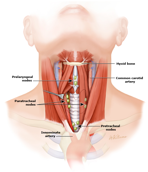
- Visceral compartment accommodates the trachea, larynx, pharynx, thyroid and parathyroid glands.
- Vascular compartment is paired and consists of the two carotid sheaths found on each side of the trachea. Each carotid sheath contains the vagus nerve, common carotid artery and internal jugular vein.
- Besides the listed structures, the neck contains cervical lymph nodes which surround the blood vessels.
Muscles and triangles
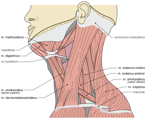
Muscles of the neck attach to the skull, hyoid bone, clavicles and the sternum. They bound the two major neck triangles; anterior and posterior.
Anterior triangle is defined by the anterior border of the sternocleidomastoid muscle, inferior edge of the mandible and the midline of the neck. It contains the stylohyoid, digastric, mylohyoid, geniohyoid, omohyoid, sternohyoid, thyrohyoid and sternothyroid muscles. These muscles are grouped as the suprahyoid and infrahyoid muscles depending if they are located superiorly or inferiorly to the hyoid bone. The suprahyoid muscles (stylohyoid, digastric, mylohyoid, geniohyoid) elevate the hyoid bone, while the infrahyoid muscles (omohyoid, sternohyoid, thyrohyoid, sternothyroid) depress it. Acting synchronously, both groups facilitate speech and swallowing.
Posterior triangle is bordered by the posterior border of the sternocleidomastoid muscle, anterior border of the trapezius muscle and the superior edge of the middle third of the clavicle. This triangle contains the sternocleidomastoid, trapezius, splenius capitis, levator scapulae, omohyoid, anterior, middle and posterior scalene muscles.
Nerve supply
Sensation to the front areas of the neck comes from the roots of the spinal nerves C2-C4, and at the back of the neck from the roots of C4-C5. In addition to nerves coming from and within the human spine, the accessory nerve and vagus nerve travel down the neck.
Blood supply and vessels
Arteries which supply the neck are common carotid arteries which bifurcate into the internal carotid artery and the external carotid artery.
Surface anatomy
The thyroid cartilage of the larynx forms a bulge in the midline of the neck called the Adam’s apple. The Adam’s apple is usually more prominent in men. Inferior to the Adam’s apple is the cricoid cartilage. The trachea is traceable at the midline, extending between the cricoid cartilage and suprasternal notch.
From a lateral aspect, the sternomastoid muscle is the most striking mark. It separates the anterior triangle of the neck from the posterior. The upper part of the anterior triangle contains the submandibular glands, which lie just below the posterior half of the mandible. The line of the common and the external carotid arteries can be marked by joining the sterno-clavicular articulation to the angle of the jaw. Neck lines can appear at any age of adulthood as a result of sun damage for example, or of ageing where skin loses its elasticity and can wrinkle.
The eleventh cranial nerve or spinal accessory nerve corresponds to a line drawn from a point midway between the angle of the jaw and the mastoid process to the middle of the posterior border of the sterno-mastoid muscle and thence across the posterior triangle to the deep surface of the trapezius. The external jugular vein can usually be seen through the skin; it runs in a line drawn from the angle of the jaw to the middle of the clavicle, and close to it are some small lymphatic glands. The anterior jugular vein is smaller, and runs down about half an inch from the middle line of the neck. The clavicle or collar-bone forms the lower limit of the neck, and laterally the outward slope of the neck to the shoulder is caused by the trapezius muscle.
