Anatomy Of The Spinal Cord
Published (updated: ).
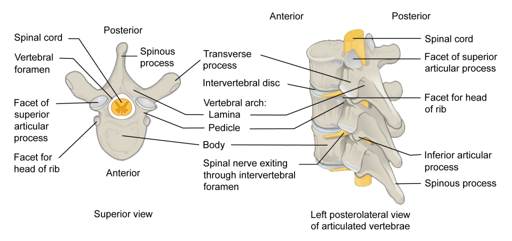
The spinal cord is part of the central nervous system (CNS), which extends caudally and is protected by the bony structures of the vertebral column. It is covered by the three membranes of the CNS, i.e., the dura mater, arachnoid and the innermost pia mater. Unlike the brain, in the spinal cord the grey matter is surrounded by the white matter at its circumference. The white matter is conventionally divided into the dorsal, dorsolateral, lateral, ventral and ventrolateral funiculi. Each half of the spinal grey matter is crescent-shaped, although the arrangement of the grey matter and its proportion to the white matter vary at different levels. The white matter gradually ceases towards the end of the spinal cord and the grey matter blends into a single mass (conus terminalis) where parallel spinal roots form the so-called cauda equina.
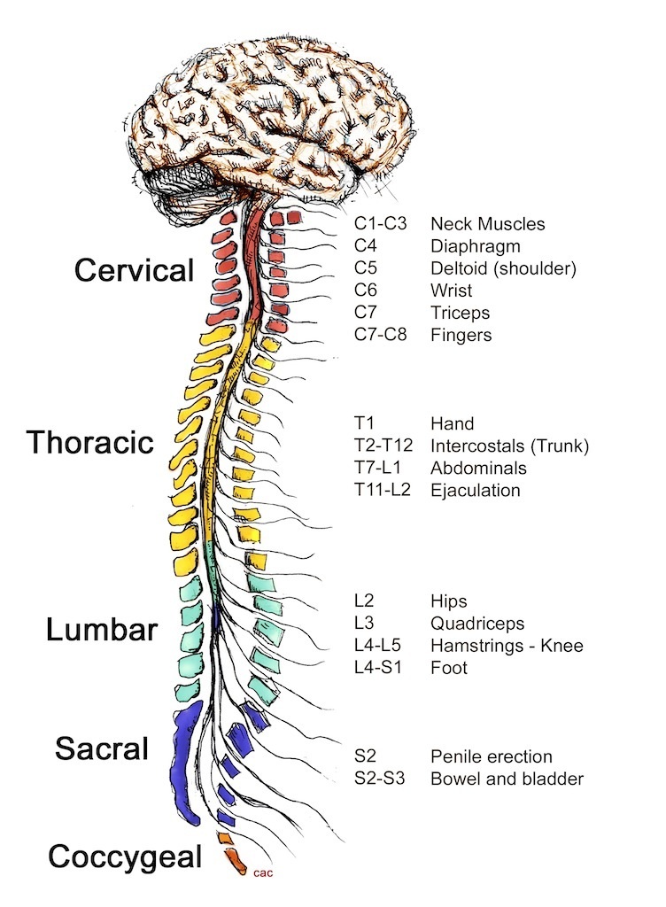
The dorsal roots leave the dorsal horn and dorsolateral white matter, coalesce into two bundles and enter the dorsal root ganglion (DRG) in the intervertebral foramen. Immediately distal to the ganglion, the dorsal and ventral roots unite and form a trunk, the spinal nerve. The spinal nerves, which are now outside the vertebral column, converge and form plexuses and from these emerge the peripheral nerves. The number of spinal nerves and spinal segments largely corresponds to the number of vertebrae with a few exceptions: there are eight cervical, 12 thoracic, five lumbar, five sacral and one coccygeal spinal segments in humans. The number of these segments varies slightly in different species.
Sensory Processing
In an oversimplified manner it can be stated that the somatic afferent functions that are processed in the spinal cord constitute the following: (a) pain and temperature, (b) touch, and (c) proprioception. Different sense organs in the peripheral structures initiate these sensory modalities, but the processing of them is usually carried out by a network of neurons in the spinal cord that are common to several of these different modalities of sensation.
Patterned Movements Organized by the Circuitry of the Spinal Cord
In addition to the monosynaptic stretch reflex the circuitry of the spinal cord can generate patterned responses that involve movement of several joints. The best explored reflex of this type is the flexor, or withdrawal reflex in response to various sensory stimuli, and in particular in response to pain. During this reflex the extremity is withdrawn from the site of the stimulus. The flexor reflex is a complex movement which involves a highly organized sequence of activation and inhibition of motoneurons to particular muscles.
The Vertebral Column
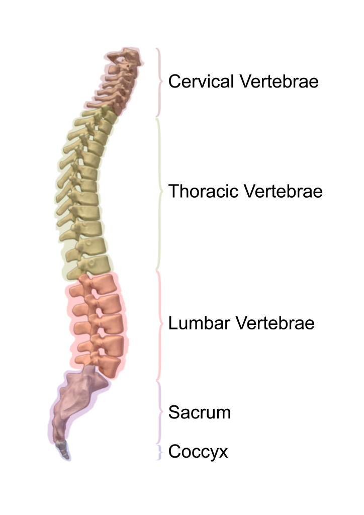
Anatomy of the spine. The spine is made up of bones, muscles, tendons, nerves, and other tissues that reach from the base of the skull near the spinal cord (clivus) to the coccyx (tailbone). The vertebrae (back bones) of the spine include the cervical spine (C1-C7), thoracic spine (T1-T12), lumbar spine (L1-L5), sacral spine (S1-S5), and the tailbone. Each vertebra is separated by a disc. The vertebrae surround and protect the spinal cord. The spinal cord is divided into segments, each containing a pair of spinal nerves that send messages between the brain and the rest of the body. Many spinal nerves extend beyond the conus medullaris (the end of the spinal cord) to form a bundle of nerves called the cauda equina.