Anatomy Of The Eye
Published .
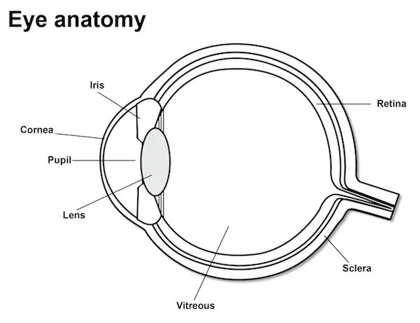
The eye is a fluid-filled sphere enclosed by three layers of tissue. Most of the outer layer is composed of a tough white fibrous tissue, the sclera. At the front of the eye, however, this opaque outer layer is transformed into the cornea, a specialized transparent tissue that permits light rays to enter the eye. The middle layer of tissue includes three distinct but continuous structures: the iris, the ciliary body, and the choroid. The iris is the colored portion of the eye that can be seen through the cornea. It contains two sets of muscles with opposing actions, which allow the size of the pupil (the opening in its center) to be adjusted under neural control. The ciliary body is a ring of tissue that encircles the lens and includes a muscular component that is important for adjusting the refractive power of the lens, and a vascular component (the so-called ciliary processes) that produces the fluid that fills the front of the eye. The choroid is composed of a rich capillary bed that serves as the main source of blood supply for the photoreceptors of the retina. Only the innermost layer of the eye, the retina, contains neurons that are sensitive to light and are capable of transmitting visual signals to central targets.
Anatomy of the human eye.
En route to the retina, light passes through the cornea, the lens, and two distinct fluid environments. The anterior chamber, the space between the lens and the cornea, is filled with aqueous humor, a clear, watery liquid that supplies nutrients to these structures as well as to the lens. Aqueous humor is produced by the ciliary processes in the posterior chamber (the region between the lens and the iris) and flows into the anterior chamber through the pupil. A specialized meshwork of cells that lies at the junction of the iris and the cornea is responsible for its uptake. Under normal conditions, the rates of aqueous humor production and uptake are in equilibrium, ensuring a constant intraocular pressure. Abnormally high levels of intraocular pressure, which occur in glaucoma, can reduce the blood supply to the eye and eventually damage retinal neurons.
The space between the back of the lens and the surface of the retina is filled with a thick, gelatinous substance called the vitreous humor, which accounts for about 80% of the volume of the eye. In addition to maintaining the shape of the eye, the vitreous humor contains phagocytic cells that remove blood and other debris that might otherwise interfere with light transmission. The housekeeping abilities of the vitreous humor are limited, however, as a large number of middle-aged and elderly individuals with vitreal “floaters” will attest. Floaters are collections of debris too large for phagocytic consumption that therefore remain to cast annoying shadows on the retina; they typically arise when the aging vitreous membrane pulls away from the overly long eyeball of myopic individuals.
