Incidence And Complications of Burns
Published .
Burn injuries are a significant problem with more than 500,000 people seeking medical treatment, 40,000 resultant hospitalizations, and 4000 deaths per year in the United States.1 The annual cost of treating these burns is estimated to be in excess of U.S. $ 1 billion, not including the indirect costs of disability and rehabilitation. These statistics have driven a multitude of studies that have systematically began to uncover the intricate mechanisms involved in burn and the complex pathophysiology of burn injury. Numerous mediators in these pathways have been the subject of animal studies in an attempt to find improved clinical therapies for treatment of burn injury. Unfortunately to date, few have translated into mainstream treatment options. Perhaps most frustrating is the lack of reproducibility of some animal studies and lack of effectiveness of potential therapies that have translated into clinical trials. On the other hand, years of clinical observations have led to a decrease in mortality following burns. This has led to a search for alternative approaches that can be used to indicate the performance of burn therapies.
A burn is one of the most critical and dangerous types of injuries that a person can suffer. The skin is the body’s largest organ, and it protects every other system of the body. If a large portion of skin is damaged, it can fail to adequately protect the heart, lungs, and circulatory system. Here are some common — but serious — complications of burn injuries.
INFECTION
A life threatening result of widespread burns across the body is sepsis, or an infection of the blood. Burned skin cannot keep harmful bacteria from the environment outside of the body, causing the body to become overrun faster than the immune system can fight them off. Sepsis can be difficult to treat and is often fatal.
LOW BODY TEMPERATURE
Skin also regulates body temperature, helping to keep critical organs like the heart and lungs warm. When a significant portion of it is damaged, it can no longer hold heat. Patients who have been burned across a large area of their bodies struggle to maintain a normal body temperature and often suffer from hypothermia, or a low body temperature. The most effective way to increase body temperature is by heating the surface of the skin — however, for a person who has been burned, even slight warmth can be utterly excruciating.
LOW BLOOD VOLUME
Burns often damage the delicate blood vessels of the body, especially when they are widespread or full thickness. This can cause blood and fluid loss, leading to a condition called hypovolemia, or low blood volume. When the body doesn’t have enough blood, organs don’t get enough oxygen. This can often lead to organ failure, especially if left untreated. Low blood volume can be treated with IV fluids and in some cases, blood transfusions.
Joint Dysfunction
Joint disorders can also be caused by burns. Damaged skin stretches tight when healing, and scar tissue can cause the abnormal stretching or twisting of limbs. Over time, this can cause joints and surrounding bones to become painful and difficult to move. In some cases, this can be permanent.
Care of burn patients

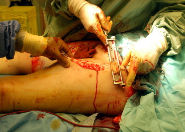
Severely burned patients are cared for on specialized intensive wards which have semi-sterile laminar flow walls, their own operating rooms, and special bathrooms for patients. After admission to hospital, the skin is often contaminated and is normally cleaned by hand in a sterile admission bath. One specific danger is that a compartment syndrome of the extremities or the trunk may develop from deep dermal burns. For example, the abdominal compartment syndrome has a mortality of over 40%. If this seems possible, a rapid escharotomy (separation of superficial burned layers of the skin) or even a fasciotomy is carried out (separation including muscle fascia).
Professional intensive care therapy is a basis for further plastic surgery therapy and plays an important role for the survival of the patient. Controlled fluid and electrolyte management with continuous and close meshed monitoring of various laboratory parameters decreases the risk of common complications of the burn injury. The most common complications are
- pneumonia (in 4.6% of all cases),
- sepsis (2.7%),
- lung failure (2.5%),
- infection of the wound (2.2%)
- acute respiratory distress syndrome (ARDS) (1.2%).
Severe complications such as cholecystitis or acute renal and organ failure must be detected early and treated adequately. Due to the necessary analgesia, patients often receive long term respiration. Therefore, the use of a tracheotomy tube is sensible.
In view of the greatly increased nutritional requirements of severely burned patients, appropriate nutrition must be initiated rapidly through a duodenal tube. The patient loses massive quantities of proteins as part of his burn injury—on the one hand through his burn wounds which release abundant quantities of protein into the bandages and on the other hand through the resulting consumption of available protein depots. Early and adequate provision of proteins not only improves the resulting osmotic gradients from intra- to the extravascular space but also the wound healing competence in affected patients. No guidelines are available for the nutrition of burned patients. Enteral food supply should be targeted as early as possible, in order to avoid regression of intestinal villi . The capillary leak, which is responsible for the massive displacement of fluids, spontaneously ceases after 24 hours. Till then, intensive fluid therapy must be continued, in order to counteract the increased cardiac output, the reduced perfusion of the kidney, the liver and the intestine, and the rapid increases in hematocrit.
Surgical therapy uses skin replacement products in the area of superficial burns. These products can be left on the injured skin until a new epithelium has been completely formed. More deeply burned areas are initially dressed in a sterile manner after cleaning and are usually treated according to the principle of early tangential excision, i.e. removal of necrotic skin (about three to four days after the accident) with wound cover as soon as possible. Different techniques must be selected according to the size, texture and thickness of the defect in the soft tissue.
On the throat, in the face, on the hands and over the joints, only cover techniques are used which later lead to satisfactory texture, color, and elasticity of the grafted skin. Thus, stigmatizing scars in visible skin areas are avoided and contractures of the scars near the joints are prevented.
Bacterial infection is a common complication which can endanger the burned patient and threaten his life. The partially damaged integrity of the skin allows devastating superficial infection which, however, is rarely the direct cause of death. In contrast, if bacteremia and consecutive sepsis develop, mortality greatly increases. 75% of patients with extensive burns die as a consequence of a severe infection. Invasive forms of infection of subcutaneous tissue layers play an especially important role, as well as surgery-related infections and superficial wound infections.
