Lower Extremity Anatomy
Published .

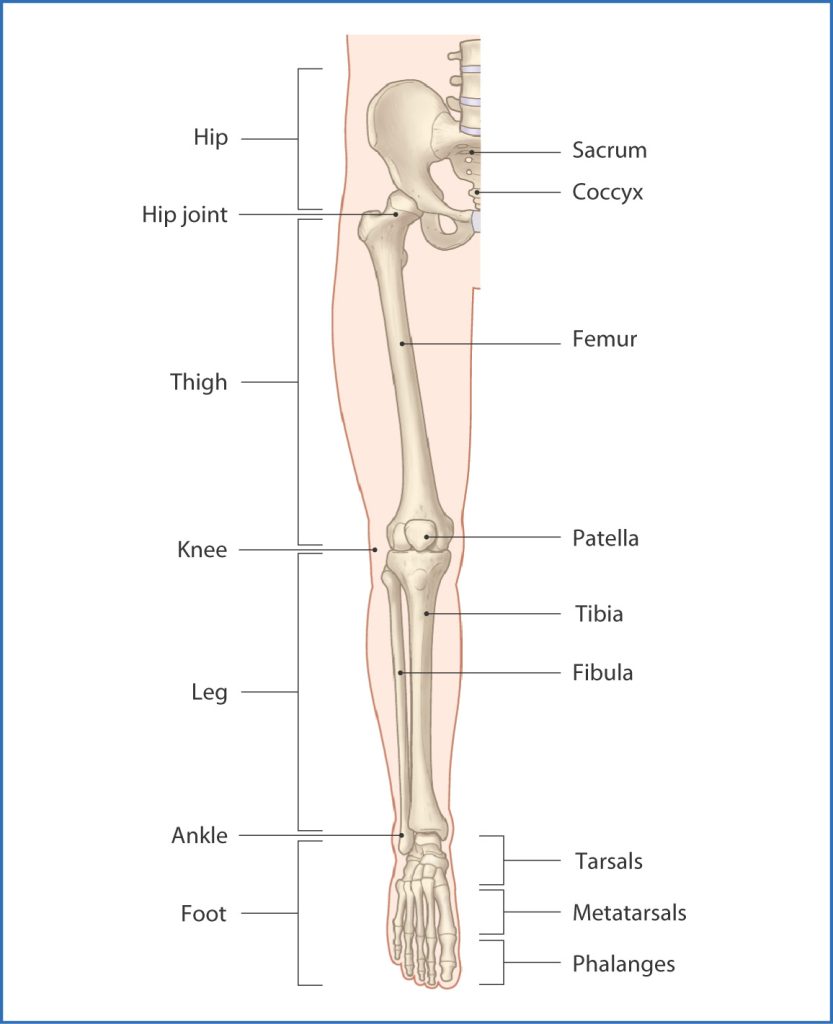
The leg is the region of the lower limb between the knee and the foot. It comprises two bones: the tibia and the fibula. The role of these two bones is to provide stability and support to the rest of the body, and through articulations with the femur and foot/ankle and the muscles attached to these bones, provide mobility and the ability to ambulate in an upright position. The tibia articulates with the femur at the knee joint. The ankle joint is a specialized articulation providing support and optimizing motion and function through the ankle joint. A normal ankle joint ultimately optimizes and allows for physiologic mobility of the foot and its associated joints and articulations.
Structure and Function
The tibia is the second largest bone in the body and provides support for a significant portion of the weight-bearing forces transmitted from the rest of the body. Proximally in cross-section, the tibia assumes a pyramidal shape/surface that articulates with the femur at the knee joint. The proximal tibia consists of medial and lateral tibial plateau surfaces, each with an associated meniscus. In the center of the two plateaus is an intercondylar spine, which contains a portion of the attachment footprint for the anterior cruciate ligament (ACL). The posterior portion contains a corresponding portion for the attachment footprint of the posterior cruciate ligament (PCL). These ligaments attach the femur to the tibia.
In addition to the ACL and PCL, knee joint stability in the coronal plane is a function of the medial collateral ligament (MCL) and the lateral collateral ligament (LCL). The MCL spans from the medial aspect of the femur to the proximal tibia distal to the joint line. The LCL attaches to the lateral aspect of the femur and courses to the anterolateral fibula head. The patellar tendon also attaches to the proximal tibia. This tendon inserts on the tibial tubercle at the midline on the tibia directly distal to the knee joint. The posterior aspect of the knee provides, in general, stability for the knee in extension. Posterior support of the knee joint is critical as the popliteal region includes the neurovascular bundle, which passes through this area to provide significant neurovascular contributions to the lower leg and foot.
The fibula is much smaller and provides much less weight-bearing support compared to its tibial counterpart. The fibula connects to the tibia via an interosseous membrane that connects the two bones distally at the ankle joint. Proximally, the proximal tibiofibular joint serves as the proximal anchoring stabilizing connection between the two bones in the lower leg. The fibula forms the lateral border of the ankle joint while the tibia forms the medial border. Relative to the ankle joint, these osseous segments are referred to as the lateral and medial malleoli, respectively.
