The Problem With Pelvic Fractures
Published (updated: ).
A pelvic fracture involves damage to the hip bones, sacrum, or coccyx – the bony structures forming the pelvic ring. Due to the inherent structural and mechanical integrity of this ring, the pelvis is a highly stable structure. Therefore, fractures of the pelvis occur most commonly in the setting of a high-impact trauma and are often associated with additional fractures or injuries elsewhere in the body.
Certain types of pelvic fractures do not disrupt the pelvic ring, such as iliac wing fractures, which can typically be managed without operative intervention. Similarly, fractures of the acetabulum are a frequent occurrence, particularly in the setting of high energy traumas, hip dislocations, and falls in the elderly, and are studied in detail classified by the anatomy of the fracture.
Although high-impact injuries are more commonly associated with pelvic fractures, they can occur in the setting of a low-impact injury as well. Low impact injuries are seen more frequently in adolescents and elderly – adolescents get pelvic fractures typically as a result of athletic injuries (e.g. avulsion fractures of superior or inferior iliac spines or apophyseal avulsion fracture of the iliac wing or ischial tuberosity), and in the elderly as a result of falls while ambulating (e.g. stable fractures of the pelvic ring or insufficiency fractures of sacrum and anterior pelvic ring). High impact injuries occur most commonly in the setting of motor vehicle accidents (e.g., vehicle collision or pedestrians struck) or fall from a significant height.
In the United States, it is estimated that pelvic fractures occur in 37 out of 100,000 individuals per year, the incidence is highest in those between the ages of 15 and 28 years. Under the age of 35, men are more commonly affected, while over 35, women are more commonly affected. Injuries to the pelvis signify a high energy mechanism of injury, and therefore a thorough trauma evaluation is needed
Pelvic ring fractures are commonly associated with injuries to the axial or appendicular spine. Therefore, the spine and extremities should also be examined assessing for limb length discrepancies and obvious angular or rotational deformities. Neurovascular structures crossing the pelvis may also be involved with injuries to the pelvis, and a thorough initial neurological examination is critical for appropriate management and monitoring.
Medical practitioners should carefully monitor the hemodynamic status of patients with pelvic fractures, as there is frequently concomitant blood loss, even in closed fractures. Intraabdominal bleeding is present in up to 40% of cases, but there also may be intrathoracic, retroperitoneal, or compartmental bleeding in such injuries. Within the pelvis, bleeding is usually caused by shearing of the venous plexus and can lead to hematomas holding up to 4L of blood. Posterior pelvic fractures may also result in an arterial injury to the superior gluteal artery, which constitutes a surgical emergency.
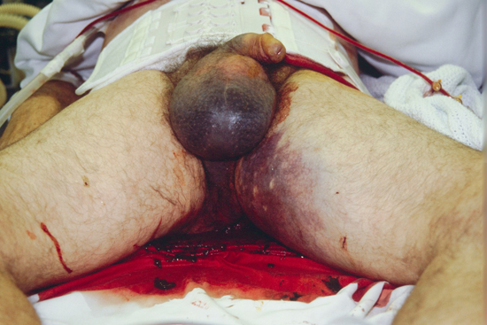
Assessment of soft-tissue injuries may provide further insight into the degree of impact sustained by the patient. It is particularly important to assess for any lacerations of the perineum (e.g., rectum or vagina) as this would indicate a severe injury and fractures potentially contaminated by urine, stool, or other environmental contaminants. Perineal ecchymosis and swelling are often signs of a pelvic fracture.
Neurologic injuries associated with pelvic fractures typically involve the L5 or S1 nerve roots. If there is a sacral fracture involved, this may also include an S2-S5 sacral nerve root injury which could result in bowel or bladder incontinence and sexual dysfunction.
Alerting features suggestive of significant pelvic injury during examination include deformity, bruising or swelling over the bony prominences, pubis, perineum or scrotum. Leg‐length discrepancy or rotational deformity of a lower limb (without fracture in that extremity) may be evident. Wounds over the pelvis or bleeding from the patient’s rectum, vagina or urethra may indicate an open pelvic fracture. Neurological abnormalities may also rarely be present in the lower limbs after a pelvic fracture. Discrete rectal or vaginal bleeding or a high‐riding prostate will not be detected in the prehospital environment.
In the alert, orientated, cooperative patient with no distracting injury, it will be possible for the prehospital practitioner to ask the patient about the presence of pain in the pelvic area, including the lower back (assessing the sacroiliac joint), groin and hips. Any positive reply should call for routine immobilisation of the pelvis. In the absence of any symptoms or signs of pelvic fracture as described above, discharge from scene is an option, provided there are no other injuries requiring transfer to a hospital. In the case of the unresponsive trauma patient, the pelvis should not be palpated for tenderness or instability.
Successful realignment of open‐book fractures with basic methods of applying a circumferential bedsheet were described in case reports of one patient in 1997 and of two patients in 2002. The authors acknowledged that the use of bed sheets was an inexact and irreproducible method. There is no control over how tightly the sheet should be applied, they are sometimes a challenge to secure with sufficient reduction force and it is not certain whether over compression of fractures could occur from using this method.
Pneumatic anti‐shock garments also known as military or medical anti‐shock trousers or G suits, have also been a popular choice for splint of pelvic fractures. However, their use restricts critical access to the abdominal and pelvic area, they are difficult to apply and do not allow for controlled pelvic reduction. Prospective randomized studies have not shown any definite benefits in reducing mortality or hospital/intensive care length of stay, and there are potential complications from their use related to compression of the lower extremities and abdomen.
An improvised use of splints, which has been suggested, is to use a Kendrick extrication device, slid under the patient upside down (with the head support towards the feet) and the straps secured around the waist and legs. This also does not use a specific amount of tension.
Prehospital use of an external pelvic compression belt device can be applied by paramedics at an accident scene within 3 seconds on clinical suspicion of unstable pelvic fractures. A variety of commercial material compression splints have been manufactured. Examples include the Stuart splint, the London splint, the Dallas pelvic binder and the Trauma Pelvic Orthotic Device (Cybertech Medical TM, California, USA). They are generally applied at the level of greater trochanters/symphysis pubis directly on to the patient’s skin.
Prehospital management of a suspected pelvic fracture should adhere to the following principles:
- Read the mechanism of injury.
- Ask the alert patient about the presence of pain in the pelvic, back or groin regions and routinely immobilize the pelvis if there is any positive reply.
- Examination is unreliable (especially if reduced GCS, or distracting injuries) and the pelvis should not be palpated, to avoid further internal hemorrhage.
- If there is any suspicion of fracture, immobilize the pelvis using an external compression splint (commercial or modified eg, sheet).
- Do not fully log roll the patient.
- Use a scoop stretcher to facilitate the patient’s movement on to a spinal board or vacuum mattress for transport. In the emergency department, this process should be reversed.
- Fluid resuscitation to maintain a radial pulse only.
- Do not remove a pelvic splint in the presence of a suspected unstable pelvic injury until it is radiologically confirmed that there is no fracture or the patient is in a theatre.
