Anatomy Of The Abdomen & Pelvis
Published .
The definition of the human abdomen is the anterior region of the trunk between the thoracic diaphragm superiorly and the pelvic brim inferiorly. Understanding the anatomy of the abdomen will ultimately serve as one’s cornerstone to understanding, diagnosing, and treating the pathology within.
Structure and Function
The abdomen ultimately serves as a cavity to house vital organs of the digestive, urinary, endocrine, exocrine, circulatory, and parts of the reproductive system.
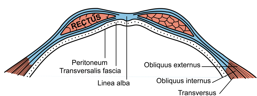
The anterior wall of the abdomen has nine layers. From outermost to innermost, they are skin, subcutaneous tissue, superficial fascia, external obliques, internal obliques, transversus abdominis, transversalis fascia, preperitoneal adipose and areolar tissue, and the peritoneum. The peritoneum is one continuous membrane; however, it is classified as either visceral (lining the organs) or parietal (lining the cavity wall). A peritoneal cavity is therefore formed and filled with extracellular fluid used to lubricate the surfaces to reduce friction. The peritoneum is comprised of a layer of simple squamous epithelial cells.
The subcutaneous tissue of the anterior abdominal wall below the umbilicus also separates into two distinct layers: the superficial fatty layer known as Camper’s fascia, and the deeper membranous layer known as Scarpa’s fascia. This membranous layer is continuous with Colles fascia within the perineal region inferiorly.
The true abdominal cavity consists of the stomach, duodenum (first part), jejunum, ileum, liver, gallbladder, the tail of the pancreas, spleen, and the transverse colon.
Blood Supply and Lymphatics
The central abdominal wall is perfused by the superior epigastric artery (branch of the internal thoracic artery) above the umbilicus, and the inferior epigastric artery (branch of the external iliac artery) below the umbilicus. Venous drainage is accomplished via the internal and lateral thoracic veins superiorly, and the superficial epigastric (branch of the femoral vein) and inferior epigastric (branch of external iliac) veins inferiorly. Lymphatic drainage above the umbilicus is accomplished mainly via the axillary lymph nodes but does drain small amounts to the parasternal lymph nodes. Below the umbilicus, lymph drains to the superficial inguinal lymph nodes.
Internally, the abdomen contains two major blood vessels – the aorta and the inferior vena cava. The aorta has three main branches that serve to supply the organs of the gastrointestinal tract, which includes the celiac, superior mesenteric, and inferior mesenteric arteries. These arteries branch off of the aorta anteriorly, while arteries that supply non-GI tract structures branch either laterally or posteriorly. Examples of such include the renal or gonadal arteries.
- The blood supply of the gastrointestinal tract follows the embryonic gut regions:
- The celiac artery supplies the foregut.
- The superior mesenteric artery supplies the midgut.
- The inferior mesenteric artery supplies the hindgut.
Note: The splenic flexure is known as a “watershed” area due to dual blood supply from distal artery branches, which can result in colonic ischemia.
Venous drainage from the organs of digestion occurs through the portal system, whereas non-digestion venous drainage occurs through the inferior vena cava and its tributaries.
The portal venous system consists of the superior mesenteric vein, inferior mesenteric vein (along with the superior rectal vein), and splenic vein and its tributaries, which all join to form the portal vein. The ligamentum teres, which contains the remnant of the umbilical vein, is of clinical significance due to its connection of the portal system to the abdominal wall. In the setting of portal hypertension, patients may experience dilation of the periumbilical veins, termed caput-medusae. Additionally, gastrointestinal cancers may metastasize to the anterior abdominal wall through the lymphatics that parallel the venous drainage, termed the Sister Mary Joseph sign/nodule.
Muscles
The abdominal muscles assist in the process of respiration, protect the inner organs, provide postural support, and serve to flex, extend, and rotate the trunk of the body.
The four main abdominal muscle groups, from innermost to outermost, can be remembered by the mnemonic TIRE: Transversus abdominis, internal oblique, rectus abdominis, and external oblique. The external and internal obliques run diagonally and perpendicular to each other. An easy way to remember which way the fibers run is to think of putting your hands in your pockets. The hand position represents the direction of the external obliques, and the internal obliques run perpendicular to this.
