Anatomy and Physiology of Organs Related To Delivery
Published .
The uterus is where the fetus lives
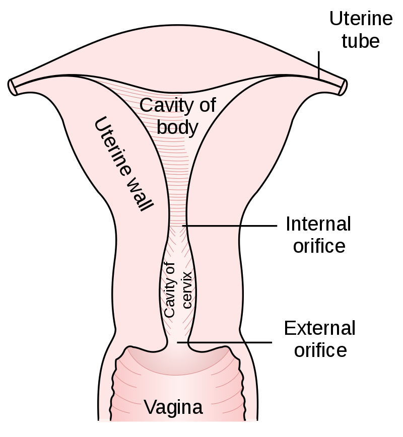
The uterus is a hollow muscular organ located in the female pelvis between the bladder and rectum. The ovaries produce the eggs that travel through the fallopian tubes. Once the egg has left the ovary it can be fertilized and implant itself in the lining of the uterus. The main function of the uterus is to nourish the developing fetus prior to birth.
The fetus is an unborn baby
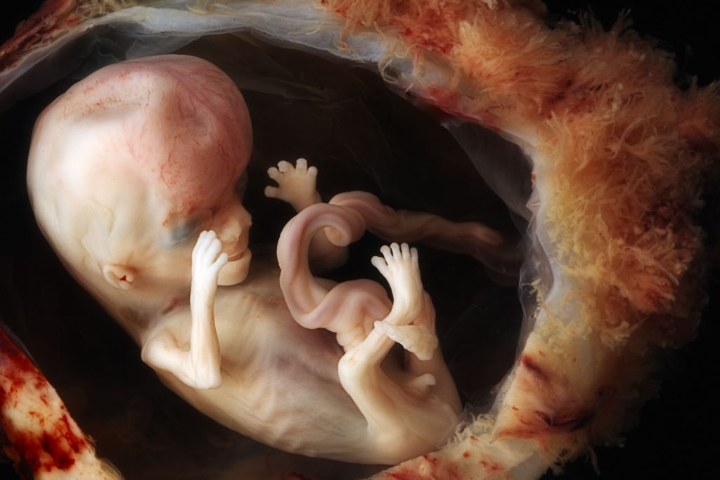
In humans, an unborn baby that develops and grows inside the uterus (womb). The fetal period begins 8 weeks after fertilization of an egg by a sperm and ends at the time of birth.
Gestation is the period of time between conception and birth when a baby grows and develops inside the mother’s womb. Because it’s impossible to know exactly when conception occurs, gestational age is measured from the first day of the mother’s last menstrual cycle to the current date. It is measured in weeks.
This means that during weeks 1 and 2 of pregnancy, a woman is not yet pregnant. This is when her body is preparing for a baby. A normal gestation lasts anywhere from 37 to 42 weeks.
The fetus is attached to the mother through the placenta
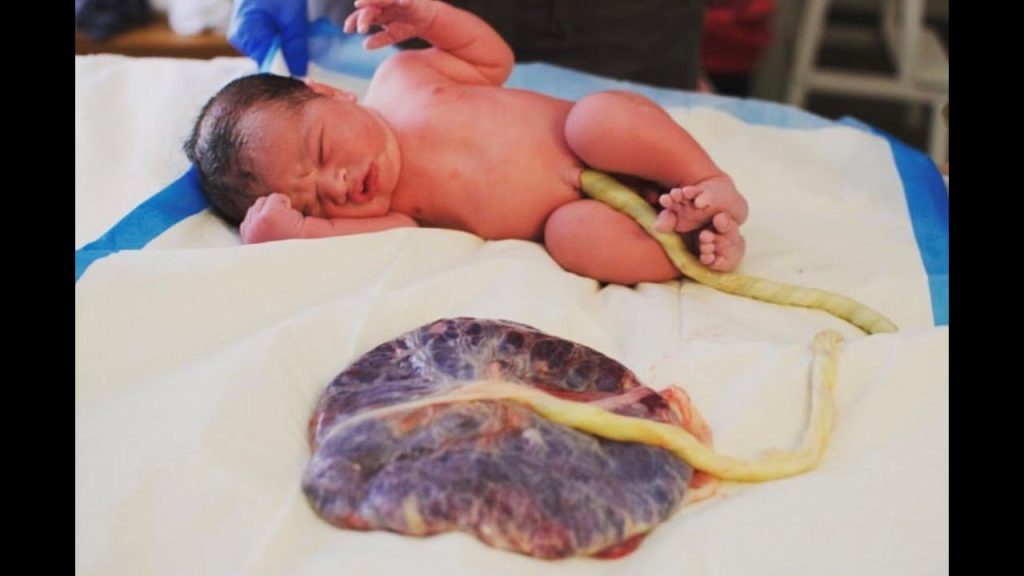
The placenta is a large organ that develops during pregnancy. It is attached to the wall of the uterus, usually at the top or side. The umbilical cord connects the placenta to the fetus.
Blood from the mother passes through the placenta, filtering oxygen, glucose and other nutrients to your baby via the umbilical cord. The placenta also filters out substances that could be harmful to the fetus and removes carbon dioxide and waste products from fetal blood.
The placenta produces a number of hormones that are needed during pregnancy, such as lactogen, estrogen and progesterone. It keeps the mother’s blood separate from the baby’s blood to protect the baby against infections. Towards the end of the pregnancy, the placenta passes on antibodies to protect the baby after birth.
The fetus is floating around in a sac filled with amniotic fluid (basically water)
While in the womb, the baby floats in the amniotic fluid. The amniotic fluid is contained in a sac called the amniotic sac. The amount of amniotic fluid is greatest at about 34 weeks (gestation) into the pregnancy, when it averages 800 mL. About 600 mL of amniotic fluid surrounds the baby at full term (40 weeks gestation).
The amniotic fluid constantly moves (circulates) as the baby swallows and “inhales” the fluid, and then releases it.
The amniotic fluid helps:
- The developing baby to move in the womb, which allows for proper bone growth
- The lungs to develop properly
- Prevents pressure on the umbilical cord
- Keep a constant temperature around the baby, protecting from heat loss
- Protect the baby from outside injury by cushioning sudden blows or movements
What does the amniotic sac not do? The amniotic sac does not carry oxygen to the lungs. The job of providing oxygenation is completed by the placenta and umbilical cord. The lungs are still developing. While in the amniotic sac contained in the uterus, the movement of the chest is primarily circulating amniotic fluid. In other words, the baby’s lungs will never take a breath until it is delivered. The pulmonary circulatory system of the fetus bypasses the lungs through the ductus arteriosus, what this means is that the blood intended for pulmonary respiration will not reach the lungs until the fetus is delivered.
When the fetus is delivered, it is squeezed through the birth canal like toothpaste
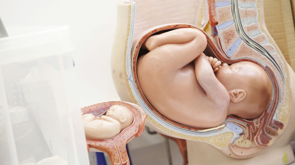
The birth canal is the muscular canal that goes from the uterus to the outside of the body. The birth canal is comprised of the uterus, cervix, and vagina, all of which are muscular enough to contract with impressive strength (commonly referred to as contractions). How strong are the contractions? Strong enough to break fingers. During birth, the baby passes through the birth canal.
What must be happening to the fetus as it progresses through the birth canal? The fetus is being compressed to 1/2 of it’s normal width. The fetus survives this intense pressure by being flexible. Fetal bones including the skull are bendable and can be compressed without causing damage to the fetus. In order to squeeze the fetus through the narrow corridor of the birth canal, parts of the birth canal become wider. The widening of the cervix (referred to as effacement) result in dislodging the mucous plug (a plug that sealed the fetus off from the outside world) out of the cervix. Once the mucous plug has been removed, the seal is broken and the pressure induced by the birth canal compress the amniotic sac until it ruptures. This is referred to as ‘breaking water’.
