Sucking Chest Wounds
Published (updated: ).
Pneumothorax is the medical term for a collapsed lung. It occurs when air enters the space around the lungs (the pleural space). This can happen when an open injury in the lung tissue causes air to leak into the pleural space. The resulting increased pressure on the outside of the lung causes it to collapse.
Pneumothorax can be traumatic or nontraumatic.
Traumatic pneumothorax results from an injury, like a blow to the chest. Nontraumatic pneumothorax can happen if the patient has lung disease, like chronic obstructive pulmonary disease (COPD), but it can also happen for no apparent reason in people without lung disease.
The long-term impact of pneumothorax can vary. If only a small amount of air is trapped in the pleural space, there may be no further complications. If the volume of air is larger or it affects the heart, it can be life-threatening.
If pneumothorax results from trauma, the symptoms often appear at the time of the injury or shortly after. Symptoms of spontaneous pneumothorax might appear when a person is at rest. A sudden attack of chest pain is often the first symptom.
Symptoms may include:
- a sudden, sharp, stabbing pain in the chest
- rapid breathing or shortness of breath (dyspnea)
- turning blue, known as cyanosis
- a rapid heart rate
- low blood pressure
- lung expansion on one side
- a hyperresonant sound when percussing the chest
- an enlarged jugular vein
- anxiety
- fatigue
Tension pneumothorax is not a classification of pneumothorax but a term that reflects the severity of pneumothorax.
- a blow to the chest
- a penetrating injury
- changes in pressure when diving, flying, or mountaineering
- a spontaneous pneumothorax progressing to a tension type
- some medical procedures
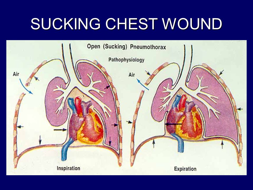
A sucking chest wound (SCW) happens when an injury causes a hole to open in the chest. SCWs are often caused by stabbing, gunshots, or other injuries that penetrate the chest.
Signs of an SCW include:
- an opening in the chest, about the size of a coin
- hissing or sucking sounds when the person inhales and exhales
- heavy bleeding from the wound
- bright red or pinkish, foaming blood around the wound
- coughing up blood
SCWs sometimes make no noise. Treat any wound caused by chest penetration as an SCW.
- Remove any loose clothing or objects covering the wound. Don’t remove clothing that’s stuck to the wound.
- Keep a hand over the wound while preparing a dressing. Protect the hand with a glove or other hand protection. If possible, have someone else put their hand over the wound. If no one else is available, have the injured person cover the wound with their hand if they’re still able to do so.
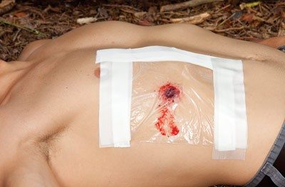
- Find a chest seal or sterile, medical-grade plastic, or tape to seal the wound. If there is no medical plastic, use a clean Ziploc bag or a credit card for the wound. EMS agencies like to use the wrapper from a petroleum gauze.
- If possible, ask the person to breathe out to release any excess air.
- Place tape, plastic, or a chest seal over any hole that’s sucking in air, including entry and exit wounds. Make sure no air enters any wound.
- Secure the tape or seal with occlusive dressingor similar wrapping material that can create a water and airtight seal. Make sure the seal has at least one open side to let out air without letting air in.
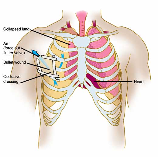
- Remove or burp the seal if the medics notice symptoms of tension pneumothorax, or a buildup of air in the chest. This happens when a lung leaks air into the chest and builds pressure. This can cause extremely low blood pressure (shock) and be fatal. Symptoms include crackling sounds when the person breathes in or out (subcutaneous emphysema), lip or finger blueness (cyanosis), enlarged neck veins (jugular vein distention), short, shallow breaths, and one side of the chest appearing larger than the other.
Keep the person on their side unless this makes it harder for them to breathe. Let out as much excess air as possible from the chest while making sure that the person can still breath.
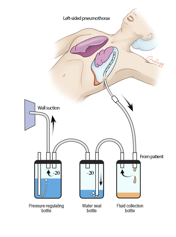
Once the person has been admitted into the hospital, surgeons may place a chest tube in the patient to evacuate air out of the pleural cavity. A chest tube is a hollow, flexible tube placed into the chest. It acts as a drain. Chest tubes drain blood, fluid, or air from around the lungs, heart, or esophagus.